

Portfolio
Educational Illustration
Scientific and Medical illustration for journal articles, textbooks, advertising, and posters.
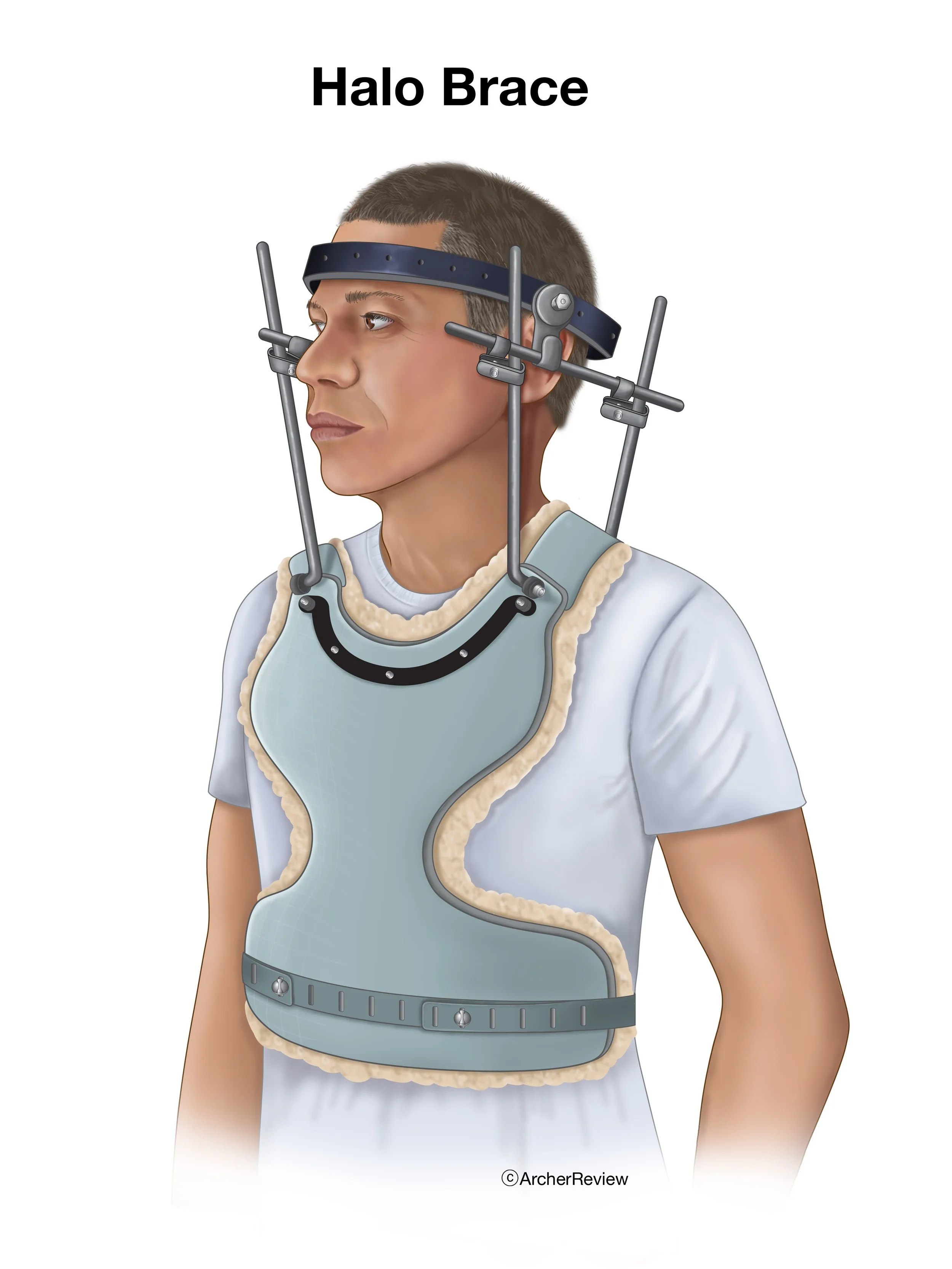
Halo Brace
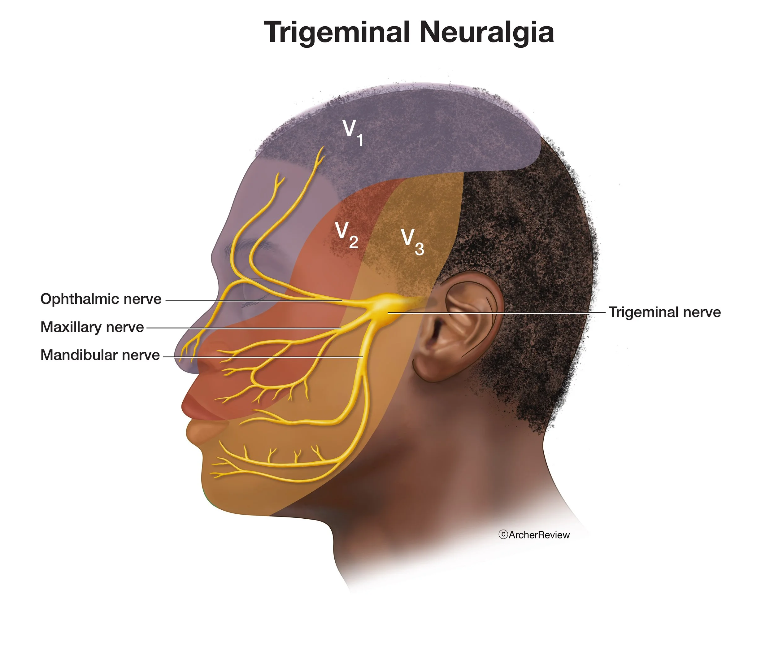
Trigeminal Neuralgia
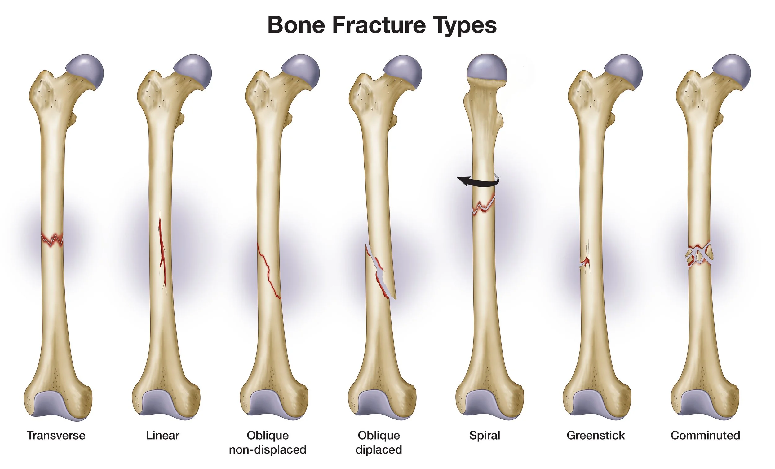
Bone Fracture Types
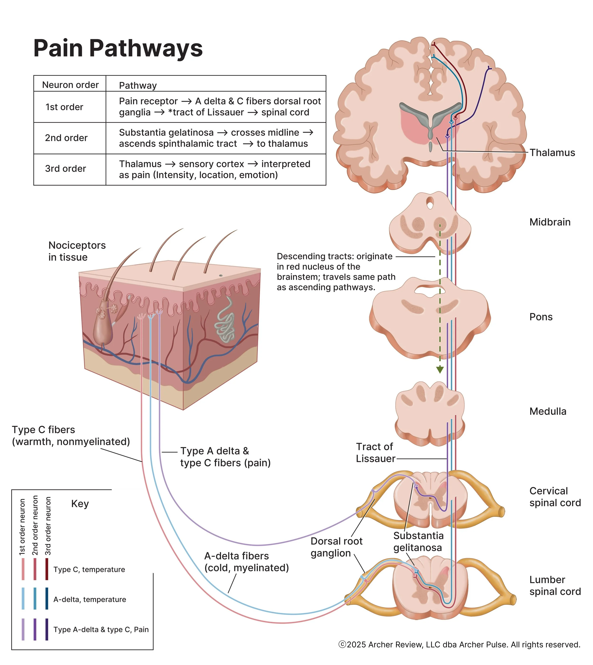
Pain Pathways
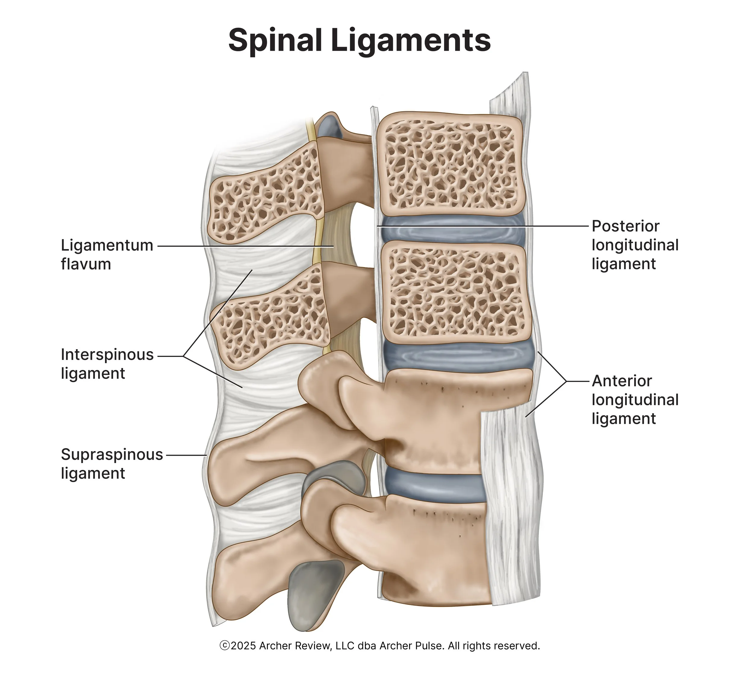
Spinal Ligaments

Fasciotomy
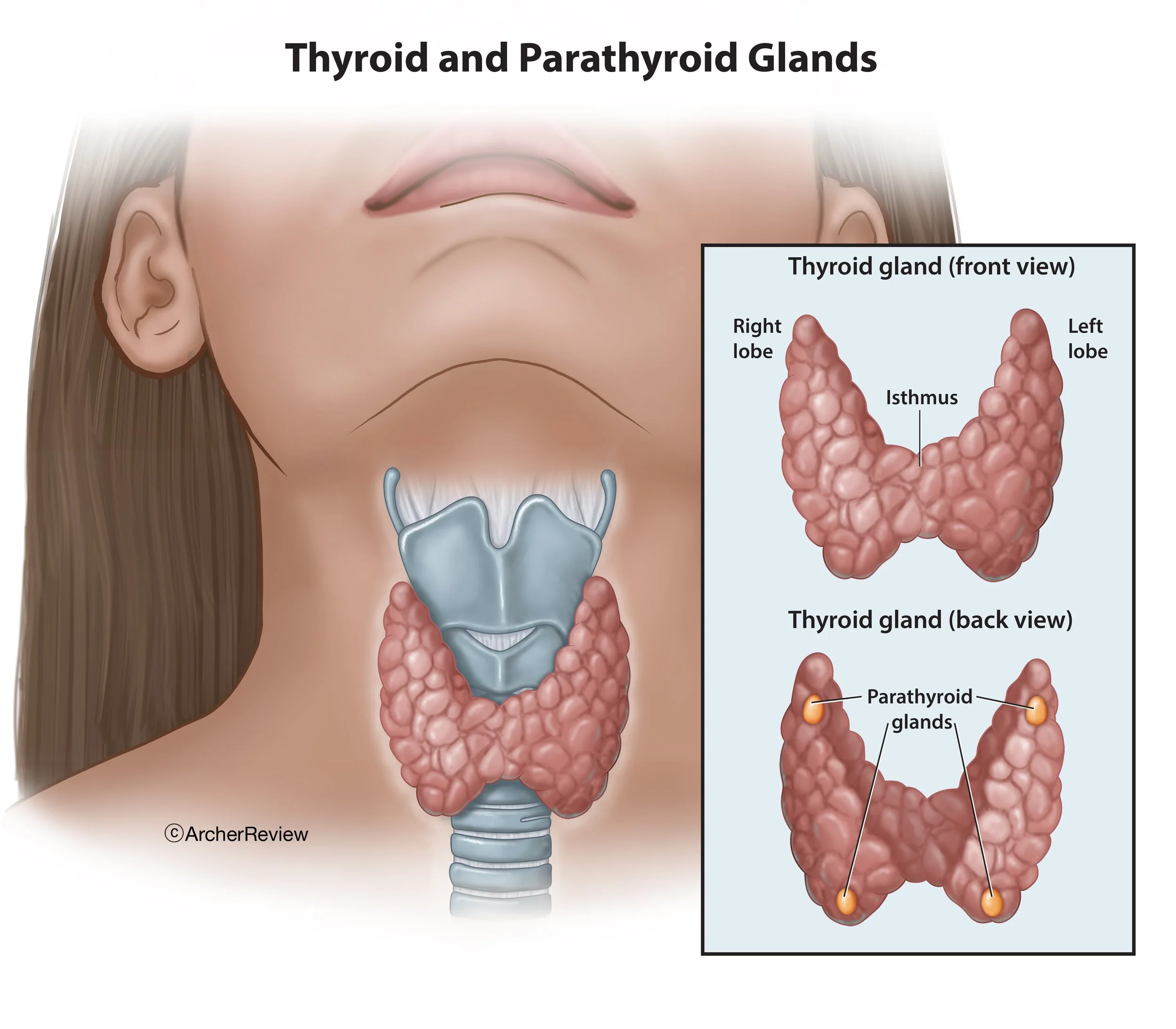
Thyroid_Parathyroid
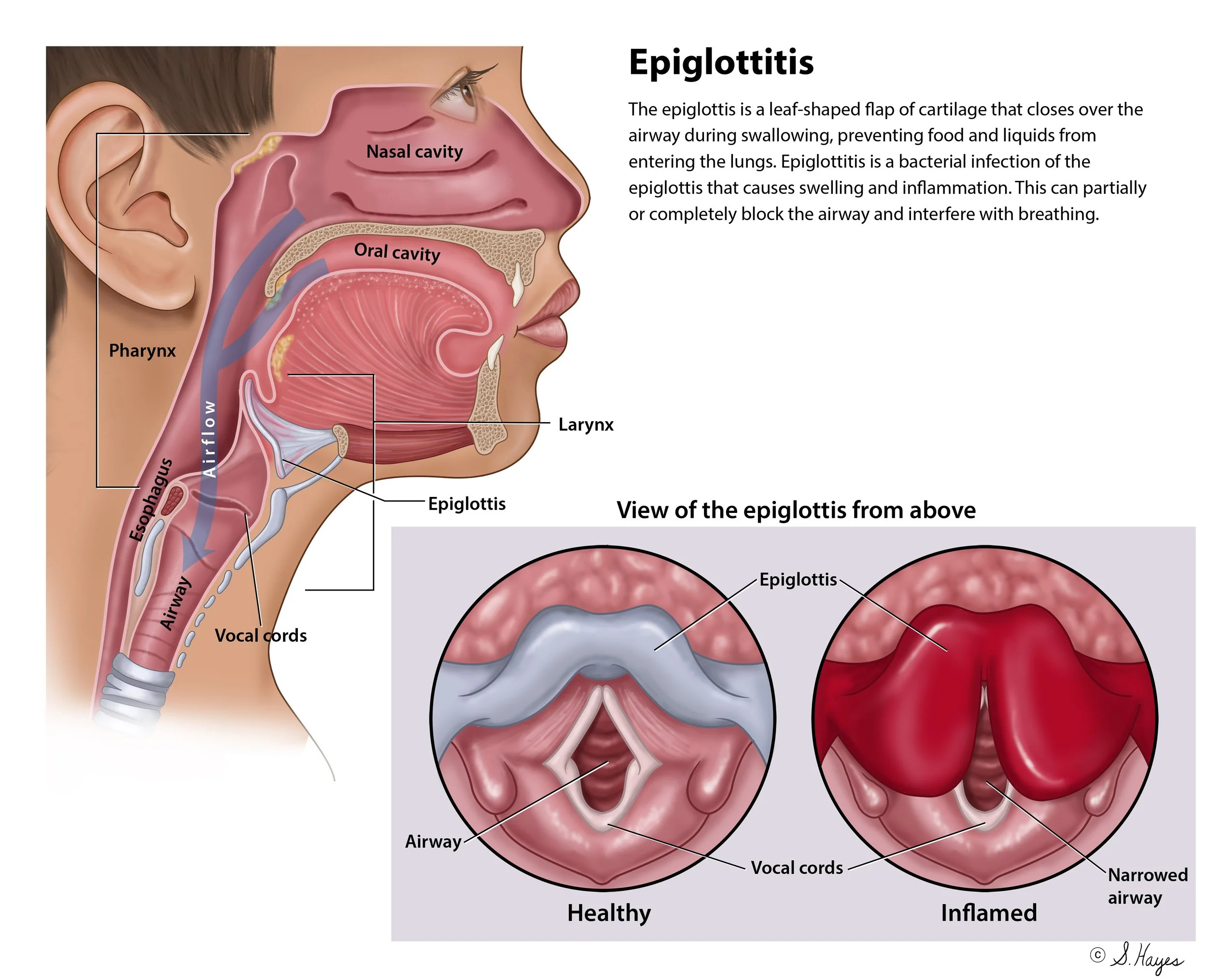
Epiglottitis
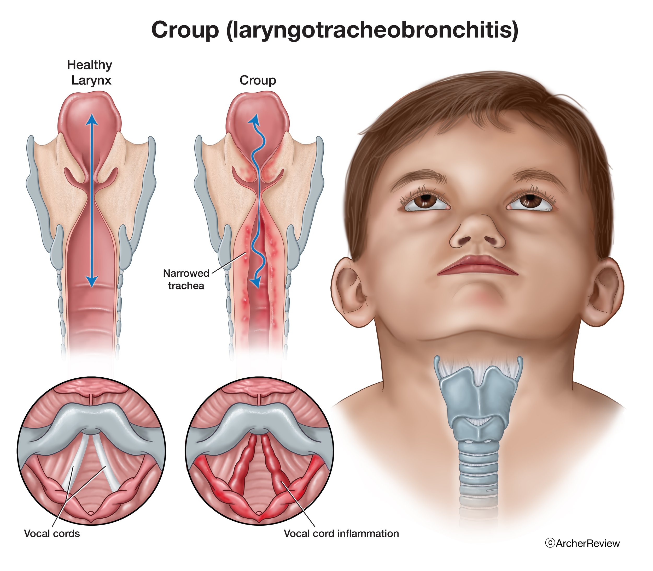
Croup (laryngotracheobronchitis)
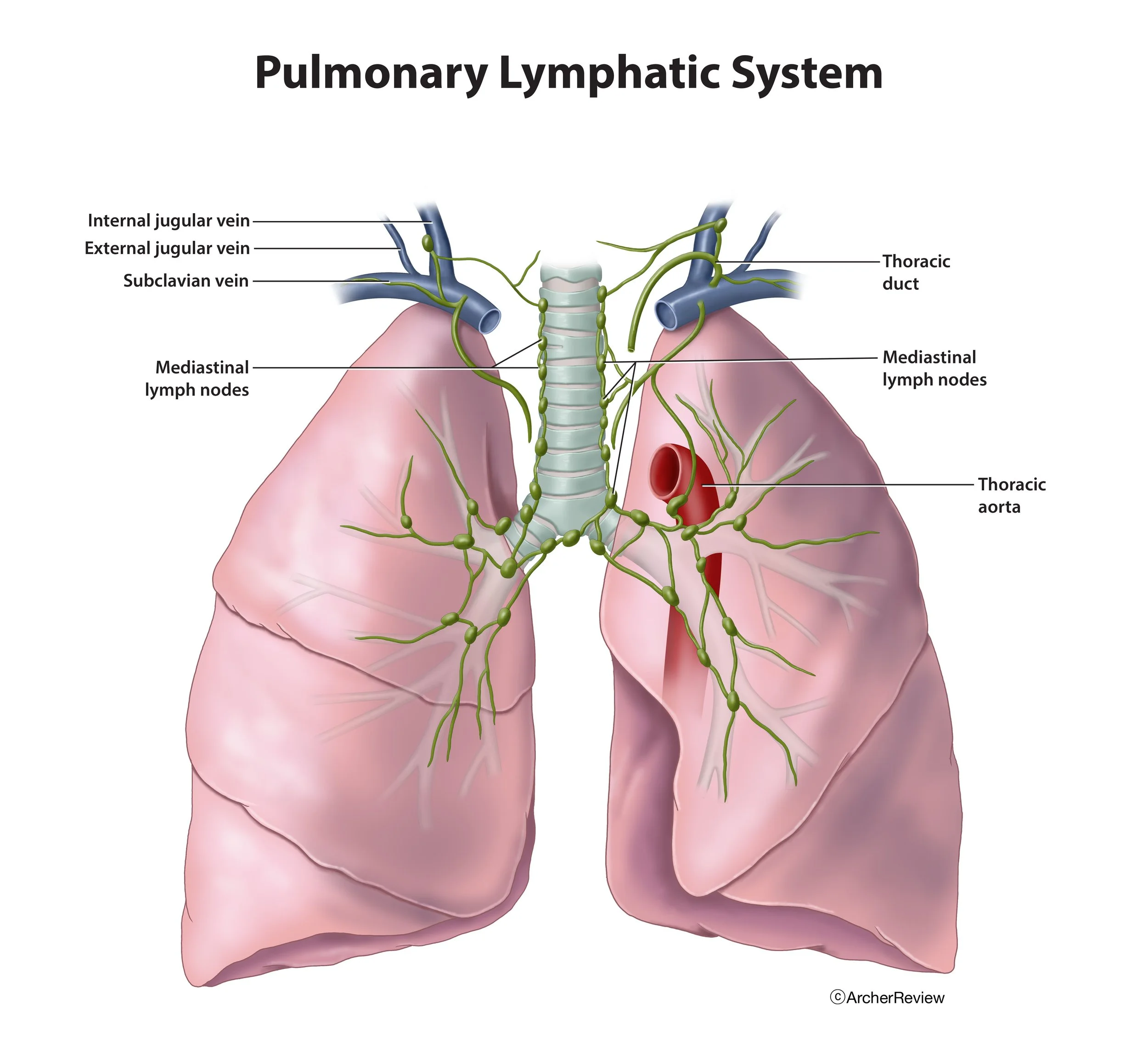
Pulmonary Lymphatic System

Anatomy of the Anus & Rectum
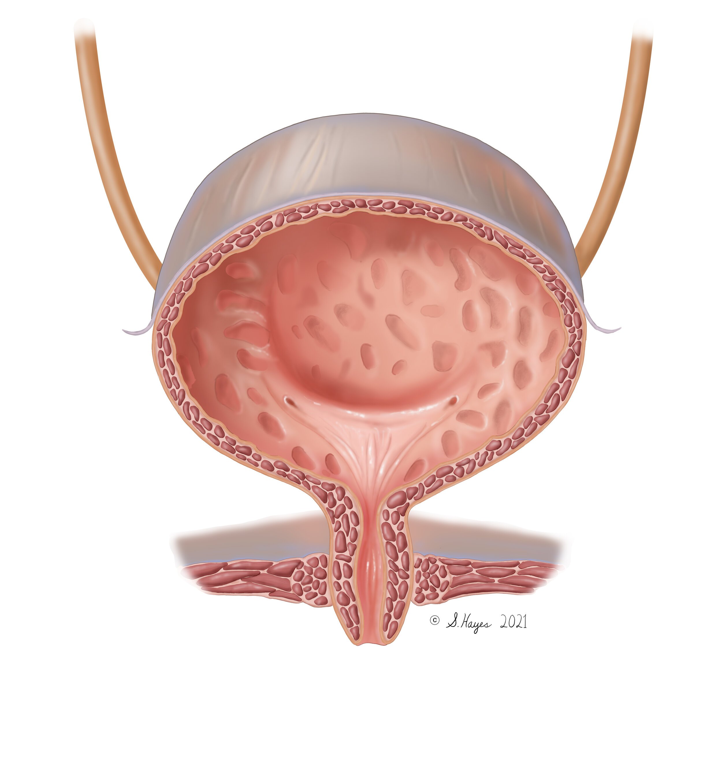
Female Urinary Bladder

Intestinal Villi

Pulmonary Hypertension

Anatomy of the Integumentary System
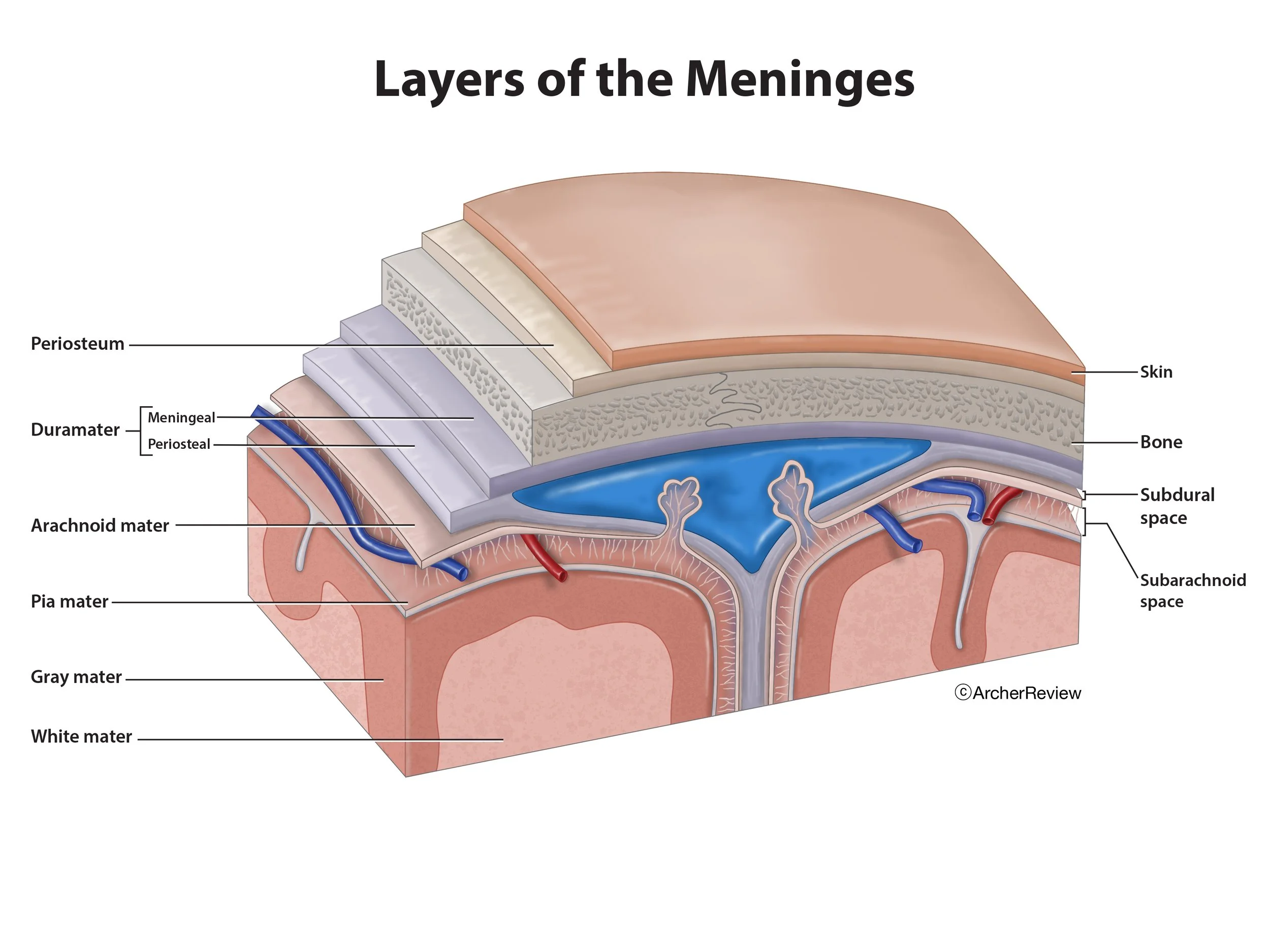
Layers of the Meninges

Burn Staging
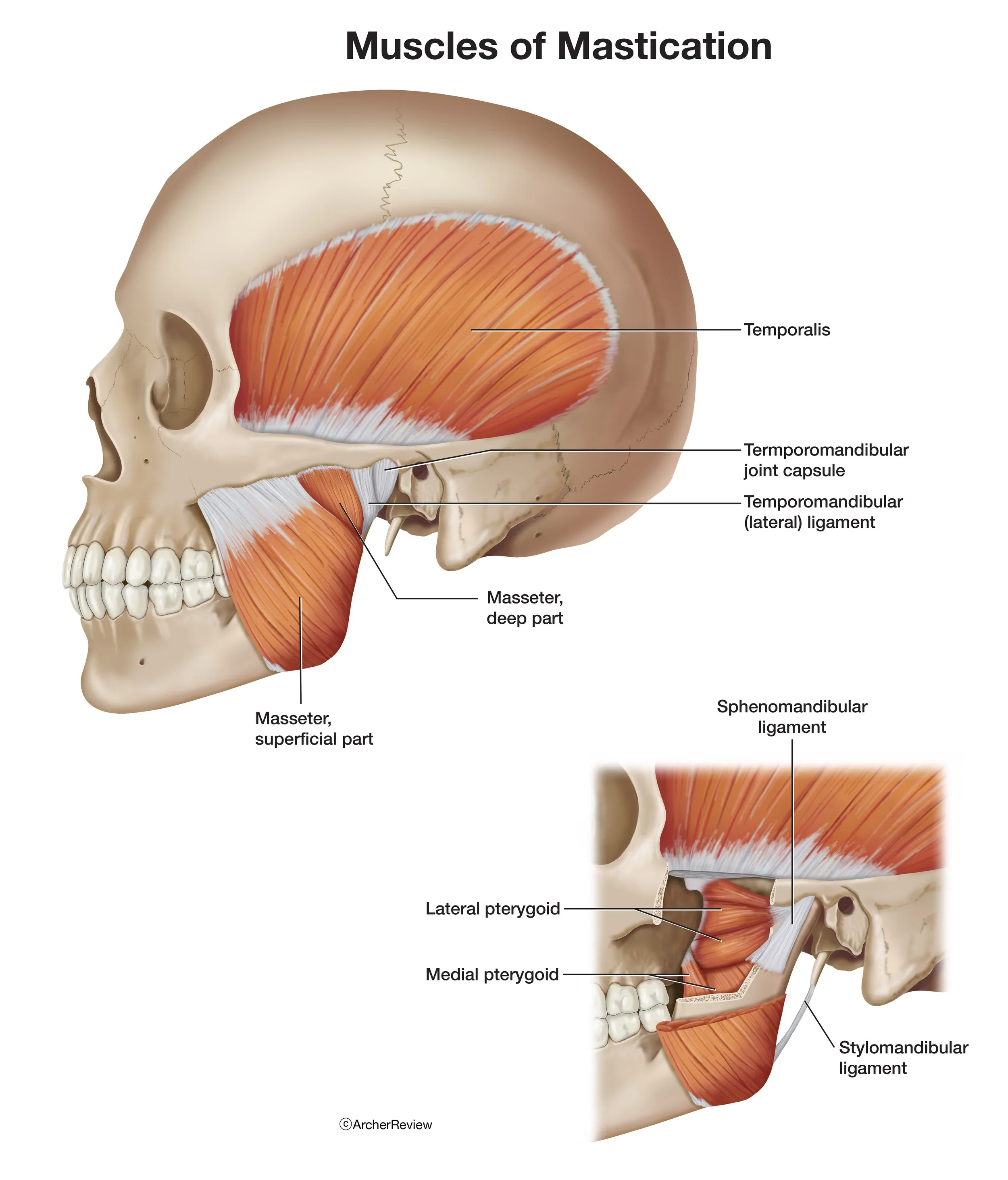
Muscles of Mastication
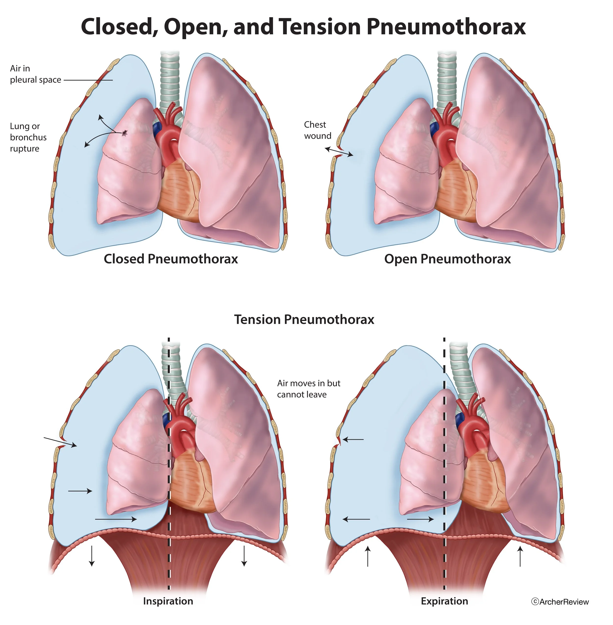
Pneumothorax

The Brain
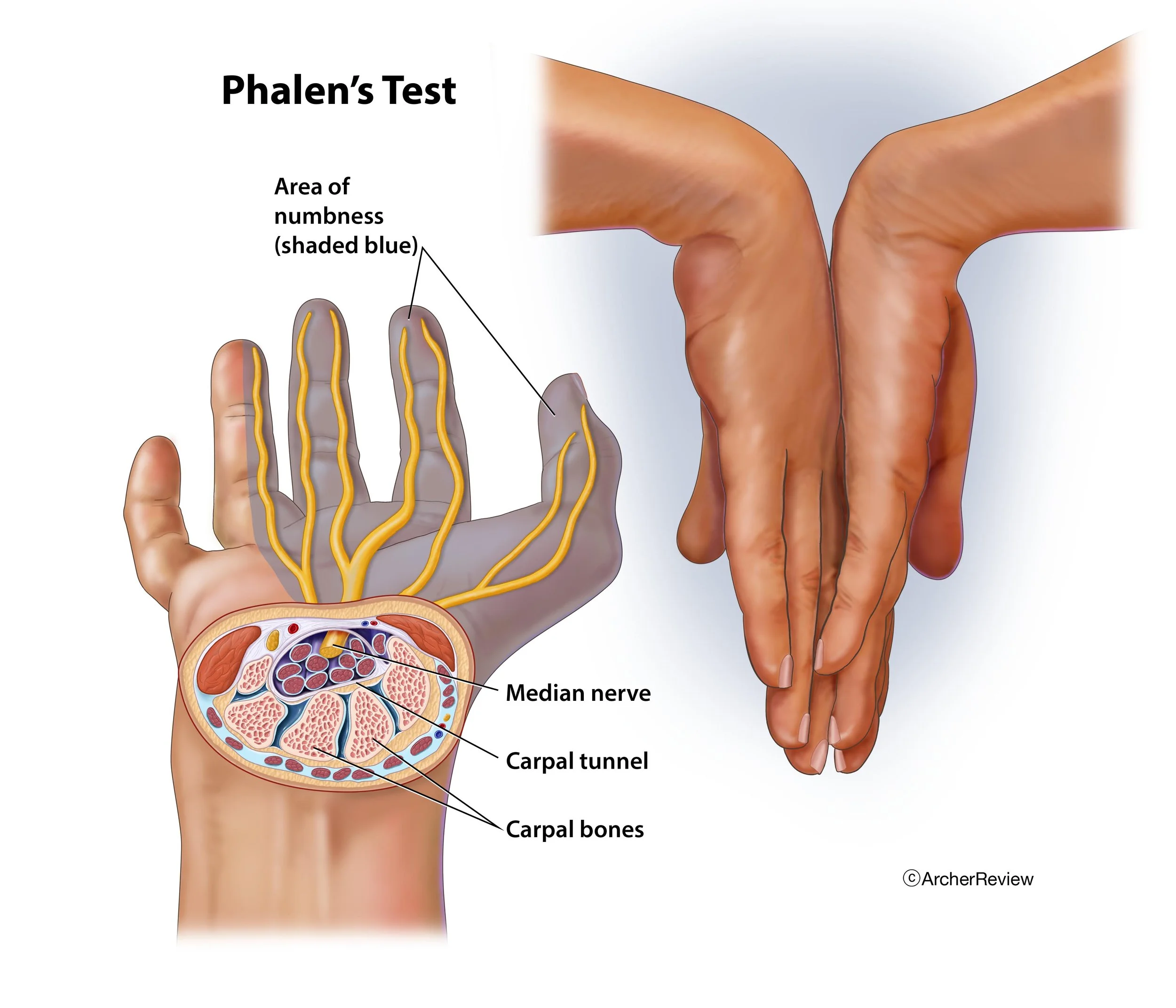
Phalen's Test

Peritoneum Structure
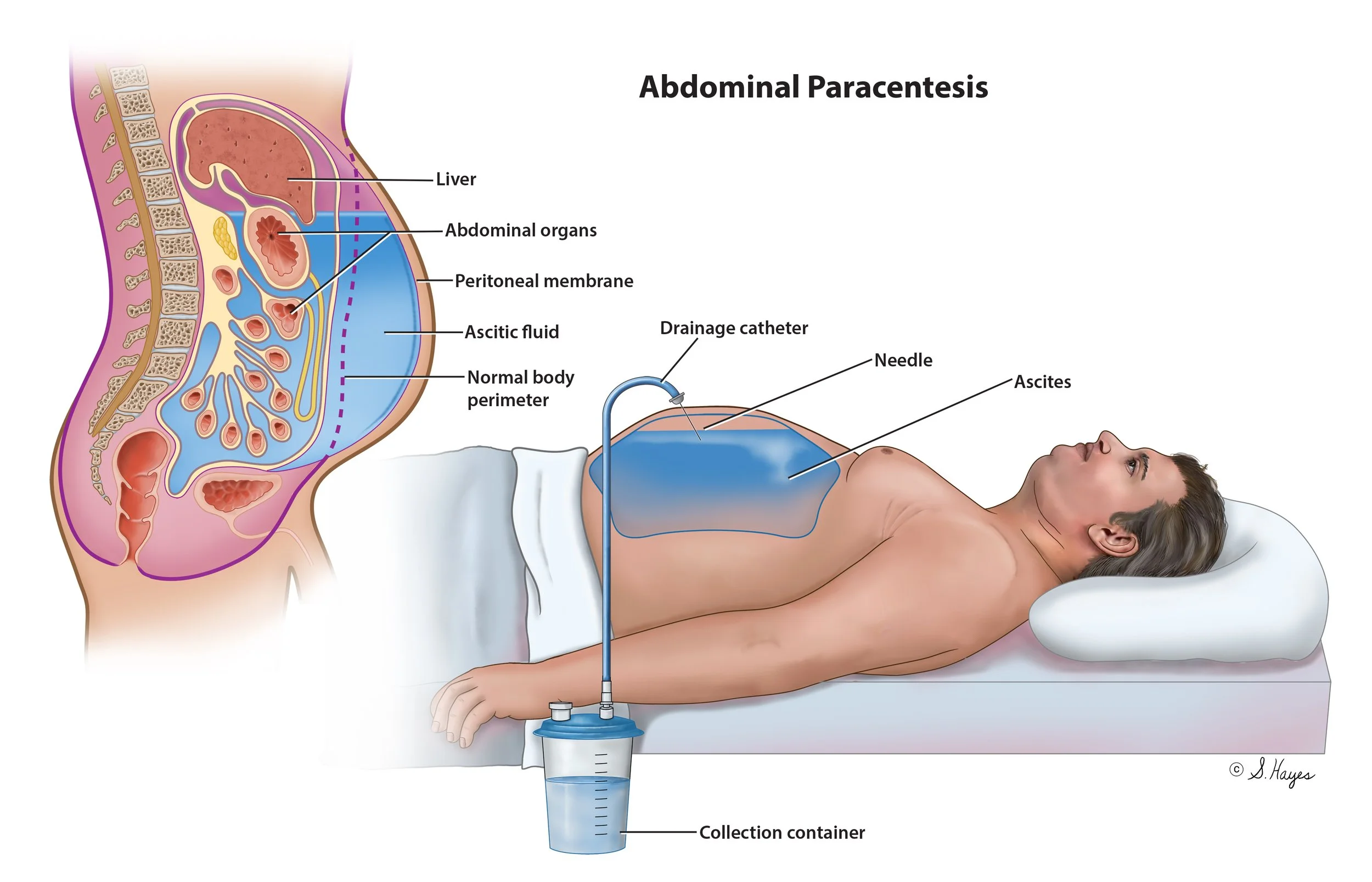
Abdominal Paracentesis

Benign Prostatic Hyperplasia

Glial Cells
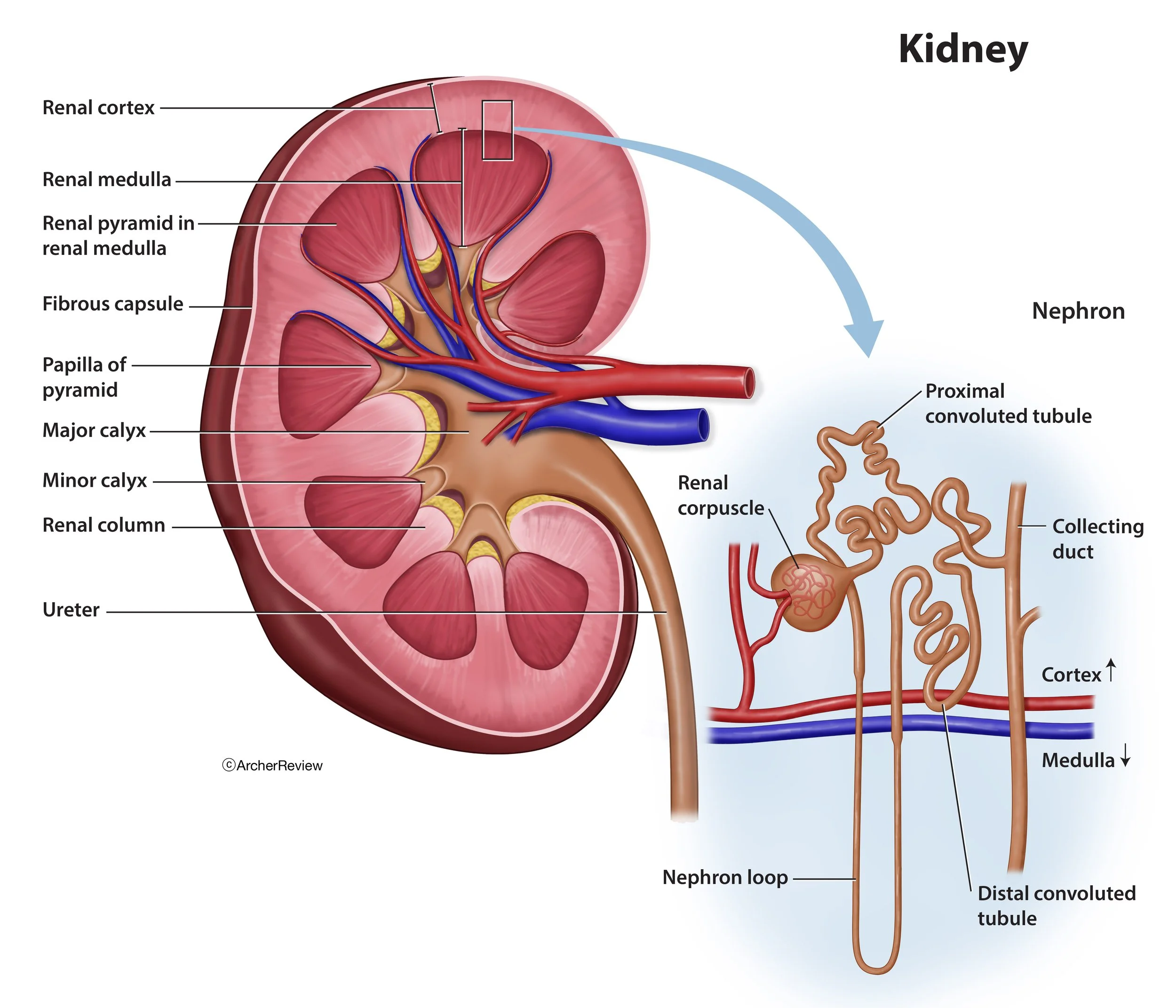
Kidney

Pyloric Stenosis

Layers of the Skin

Gall Bladder

Salivary Glands

Stages of Caries Development
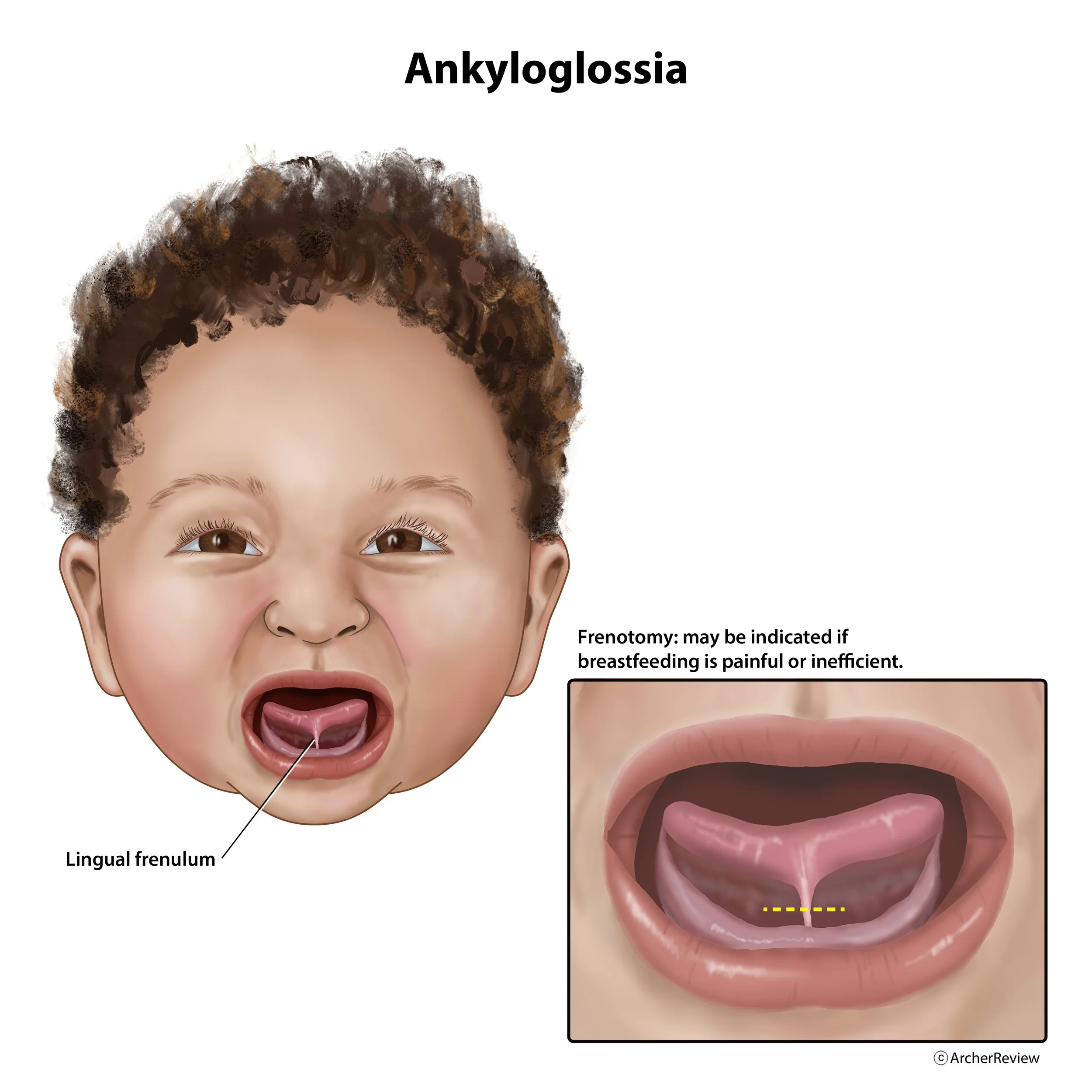
Ankyloglossia

Pharynx/Esophagus
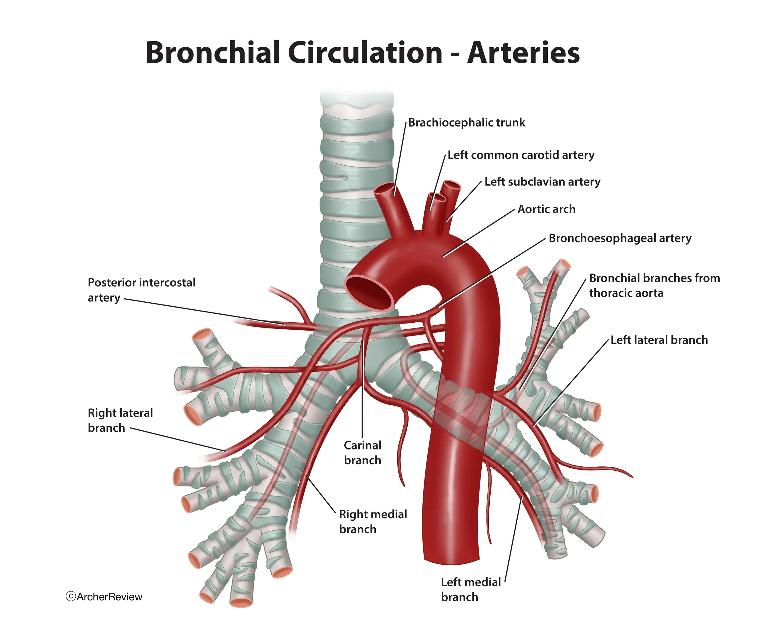
Bronchial Circulation - Arteries
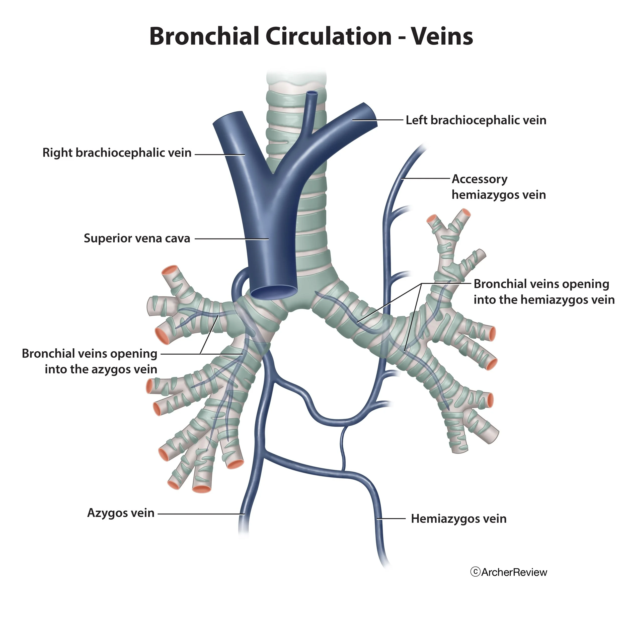
Bronchial Circulation - Veins

Hyphema

The Spinal Cord

Glaucoma

Placenta Previa

Pulmonary Embolism
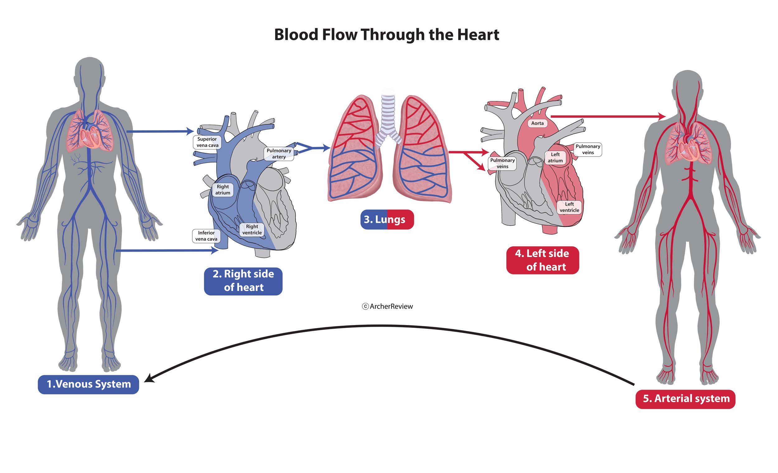
Blood Flow Through the Heart
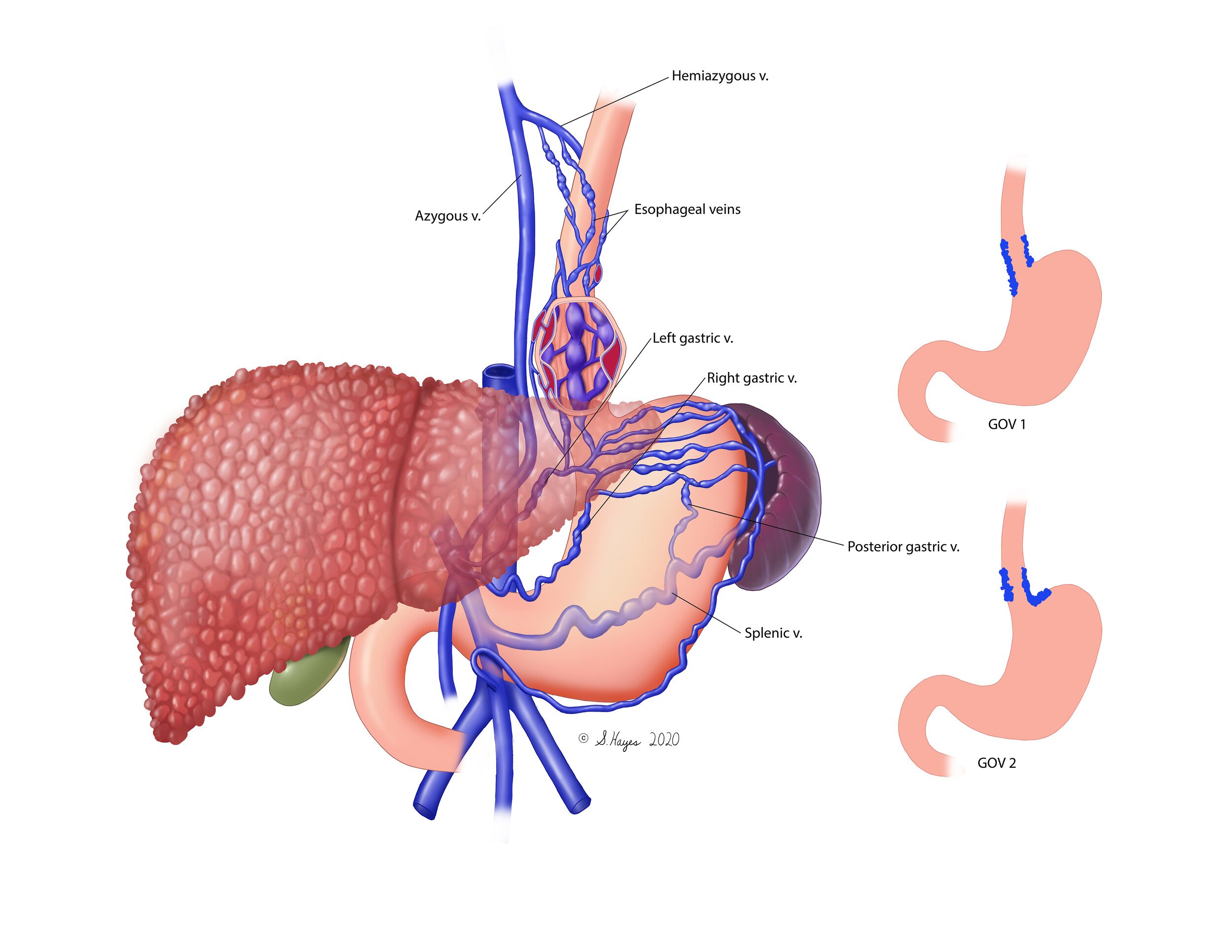
Gastroespohageal Varices
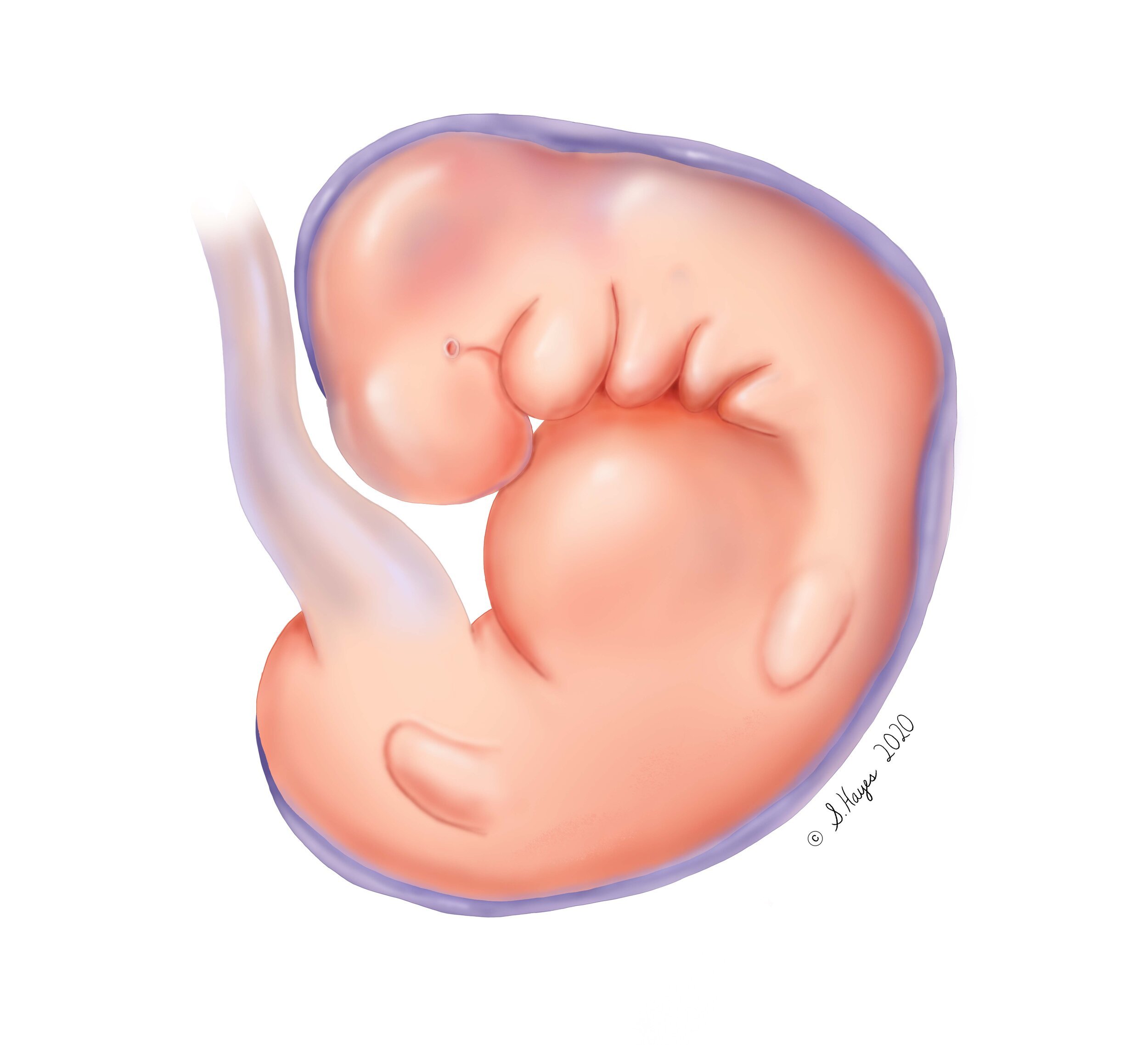
Embryo

Cerebrospinal Fluid Circulation

Cardiac Cycle
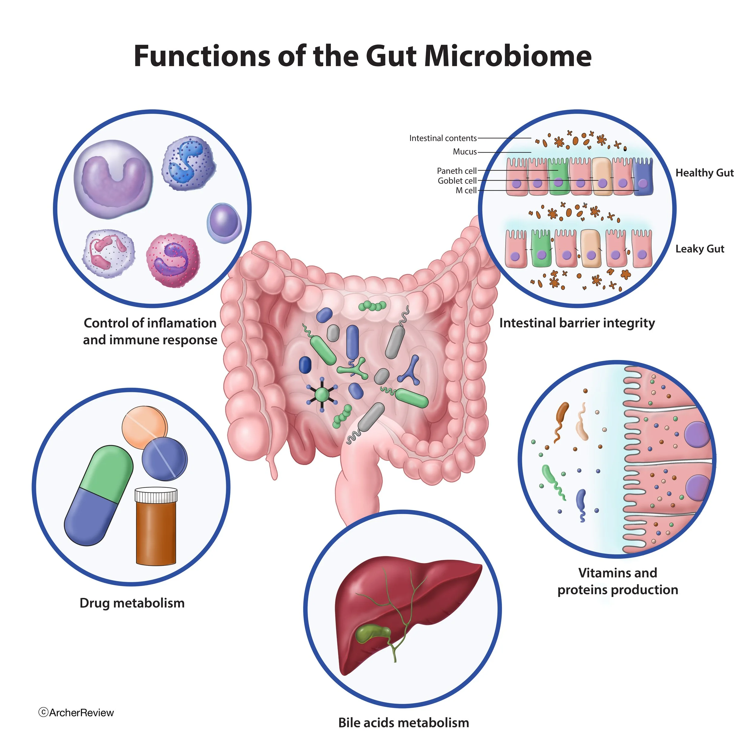
Functions of the Gut Microbiome

Stasis Dermatitis

vascular plug
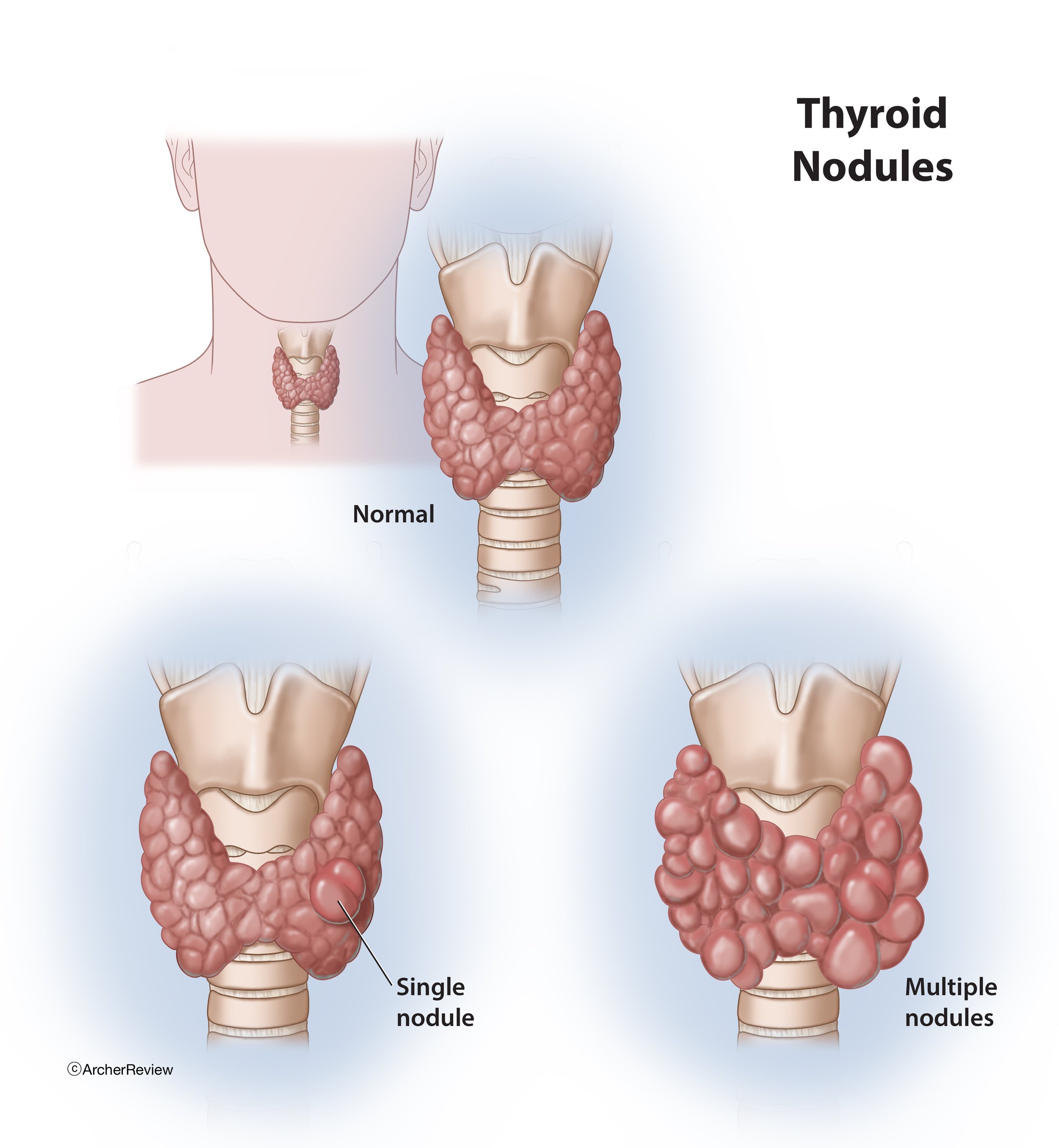
Thyroid Nodules

Illustration of a knee joint with a highlighted area indicating pain or injury, showing bones, cartilage, and soft tissue.

An illustration of the inside of a human bone showing the marrow, blood vessels, and spongy bone structure.

Types of Bone Cells
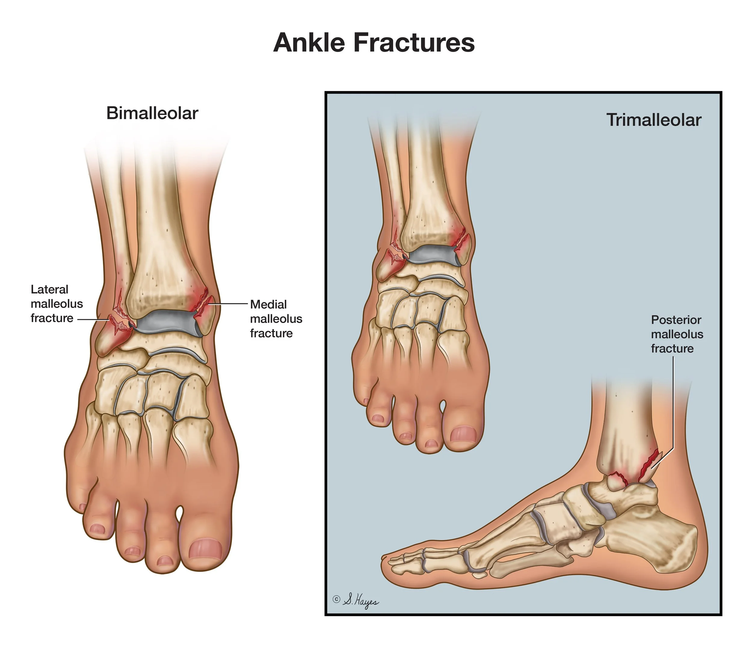
Ankle Fractures

Breast Anatomy

Creating a Sterile Field

Micrognathia

Diseased Liver

Epididymitis

Pertussis

Finkelstein Test

Pancreas

Liver Anatomy
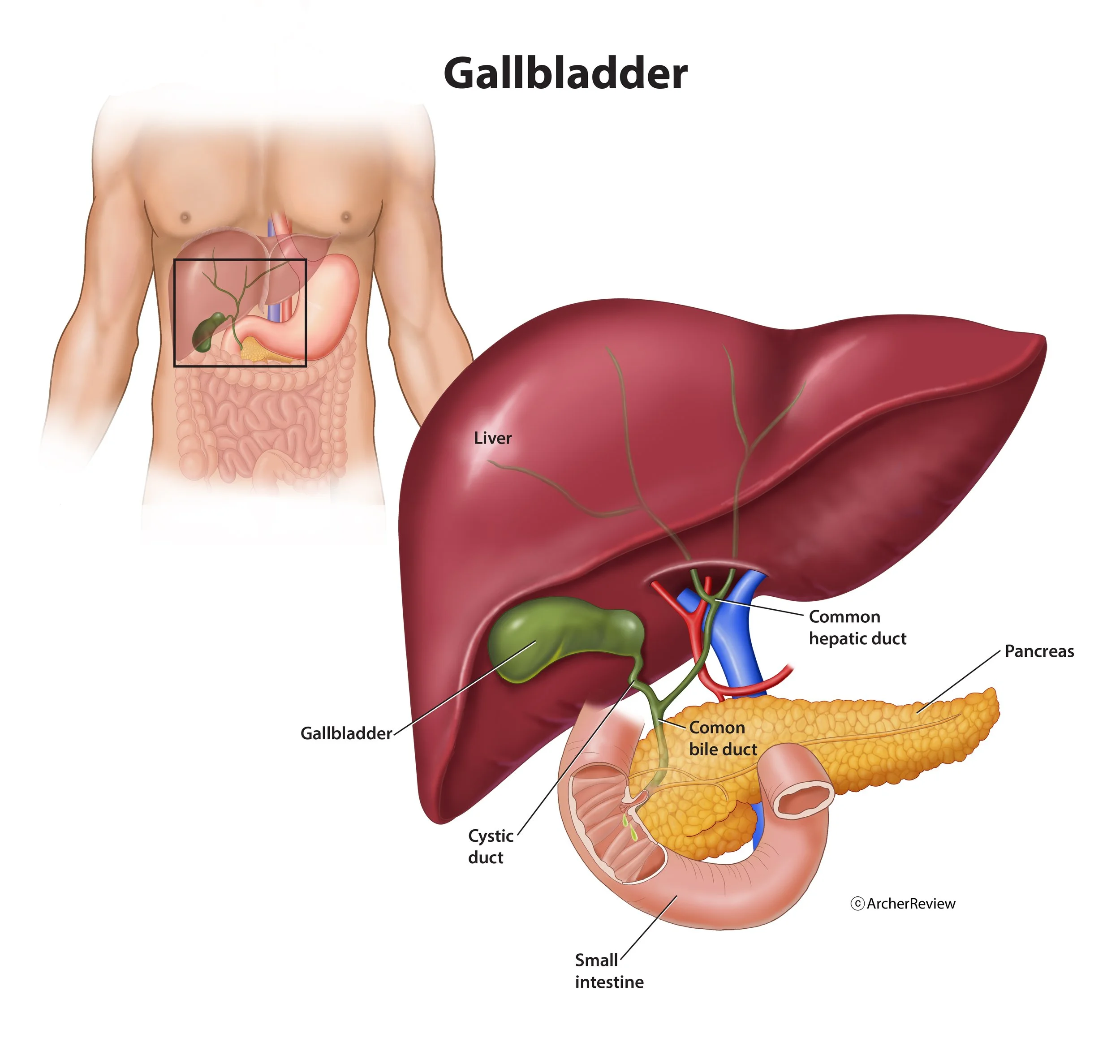
Gallbladder
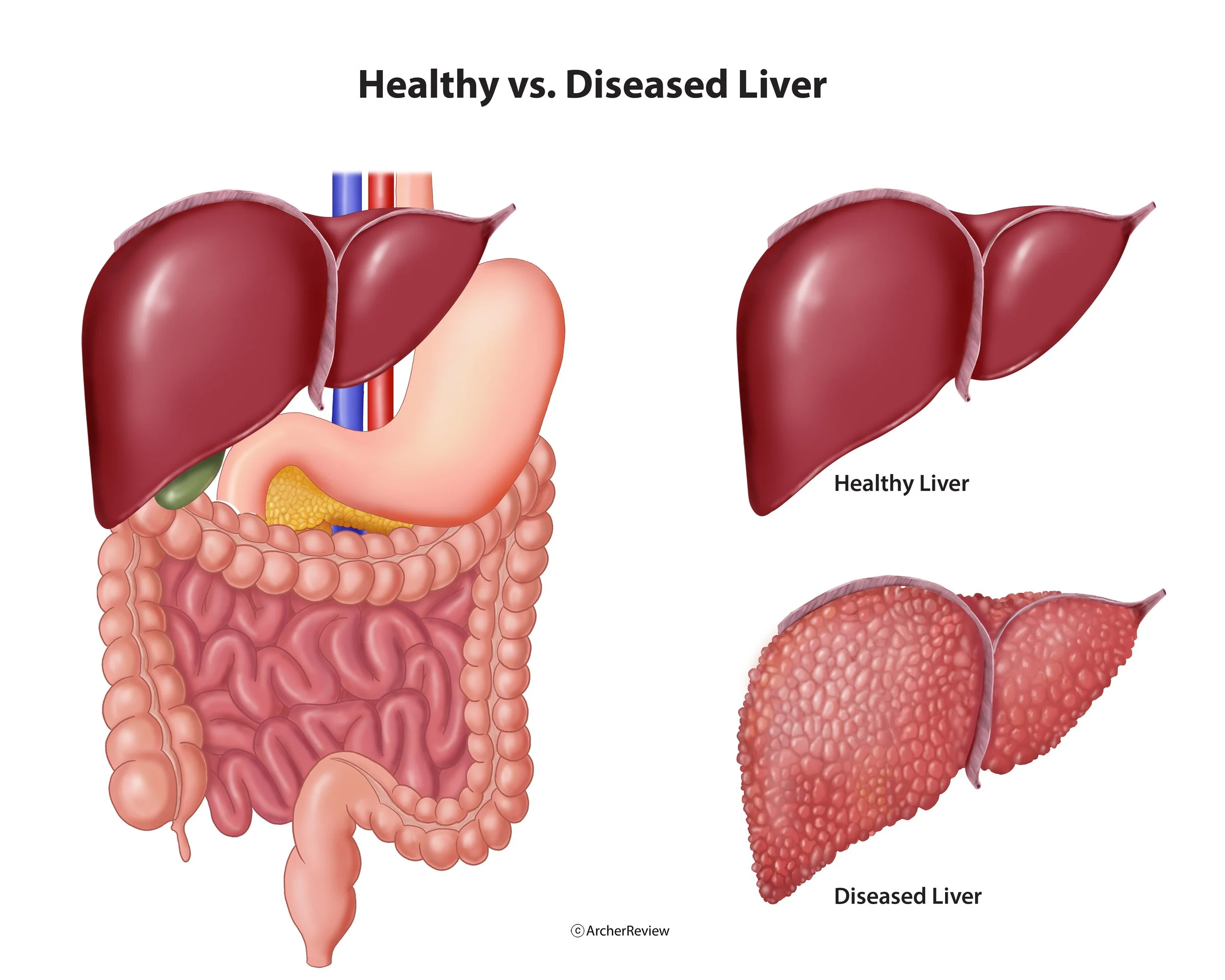
Healthy vs Diseased Liver
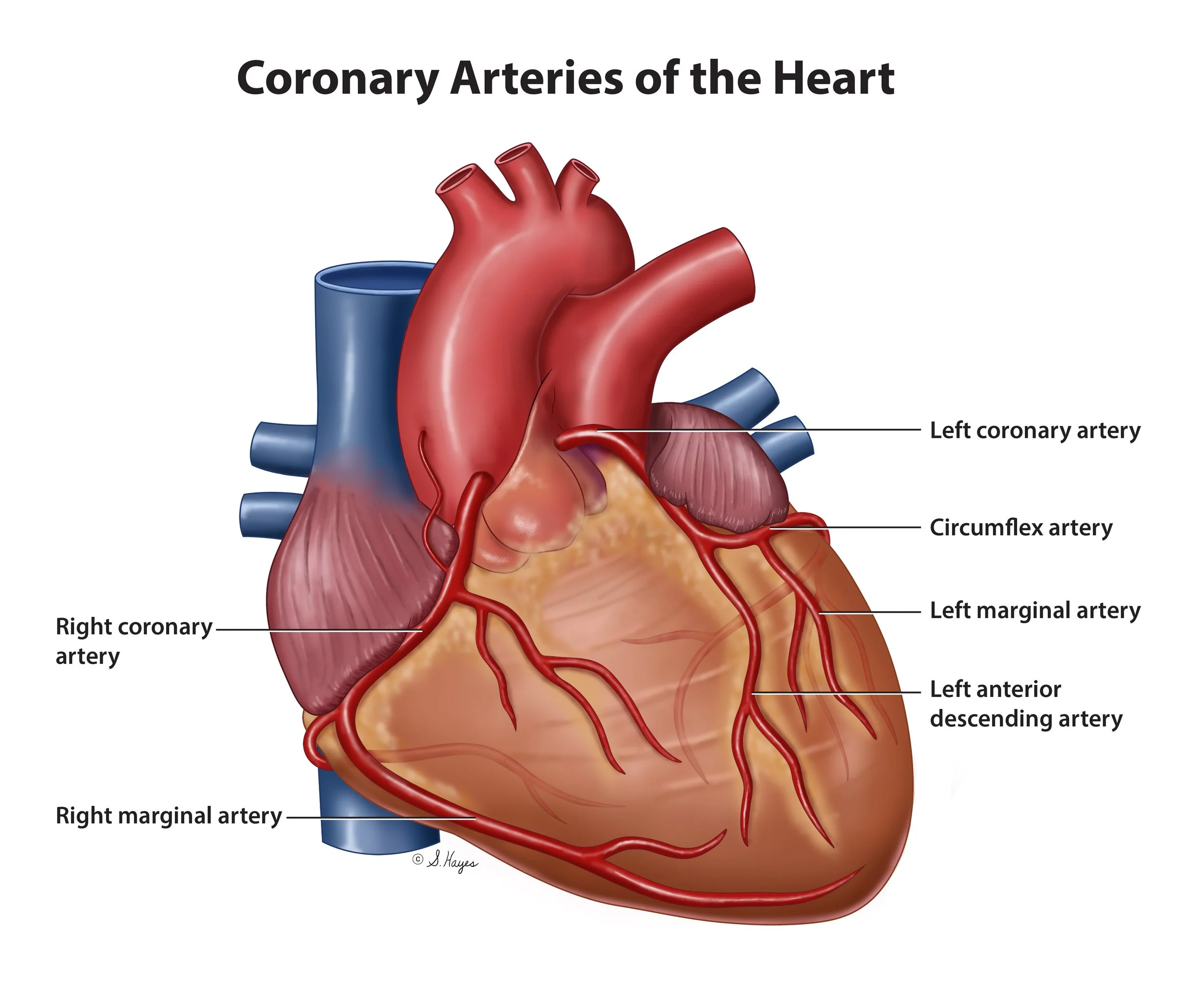
Coronary Arteries of the Heart

Heart Anatomy

Leff-Calve-Perthes Disease

Lobes of the Brain

Muscle Fiber Anatomy
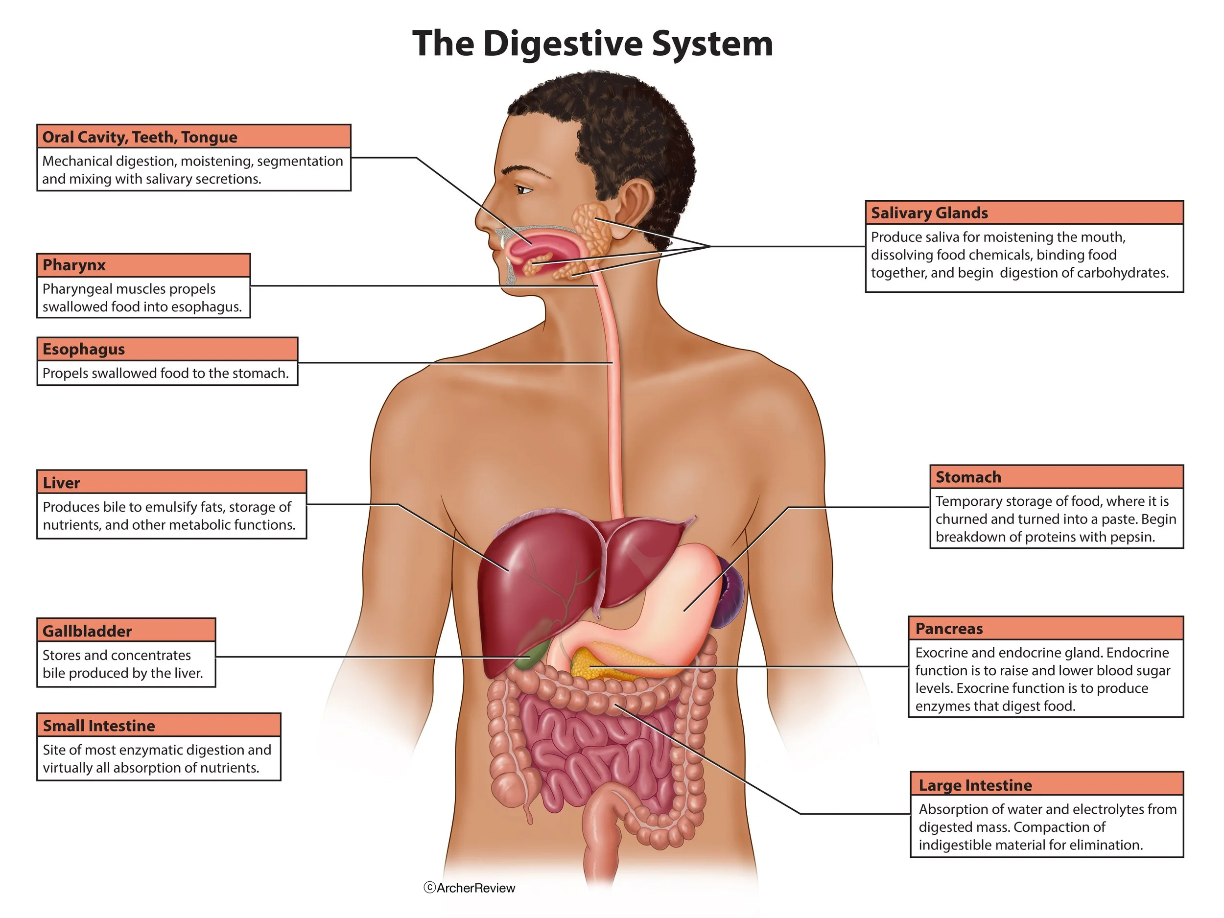
The Digestive System

Peritoneal Dialysis
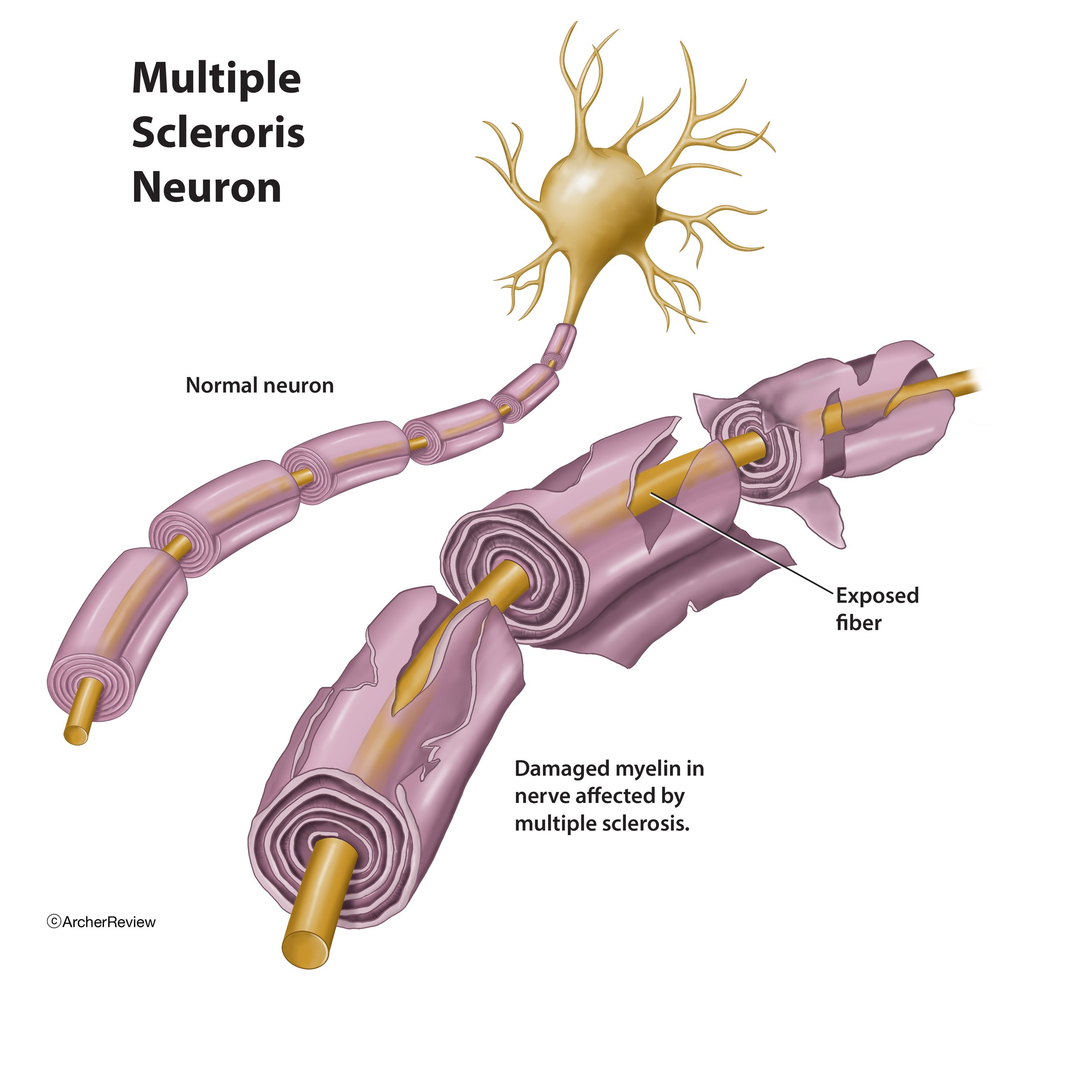
Multiple Sclerosis Neuron
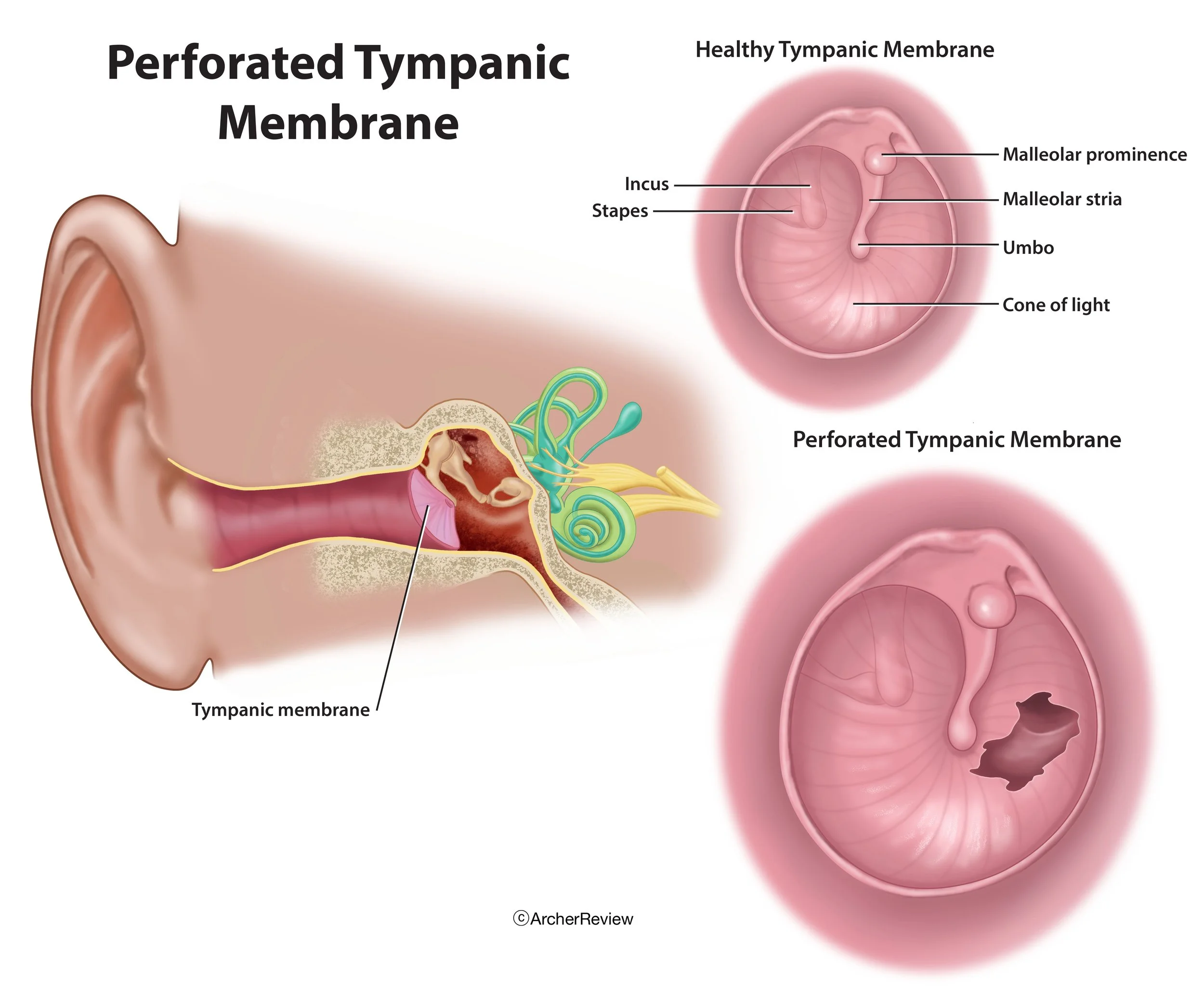
Perforated Tympanic Membrane

Meniere's Disease
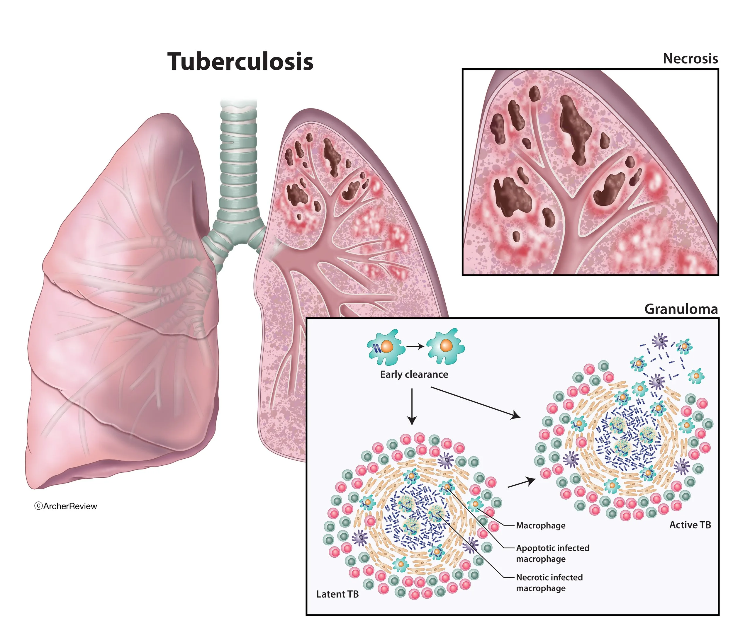
Tuberculosis

Testicular Torsion
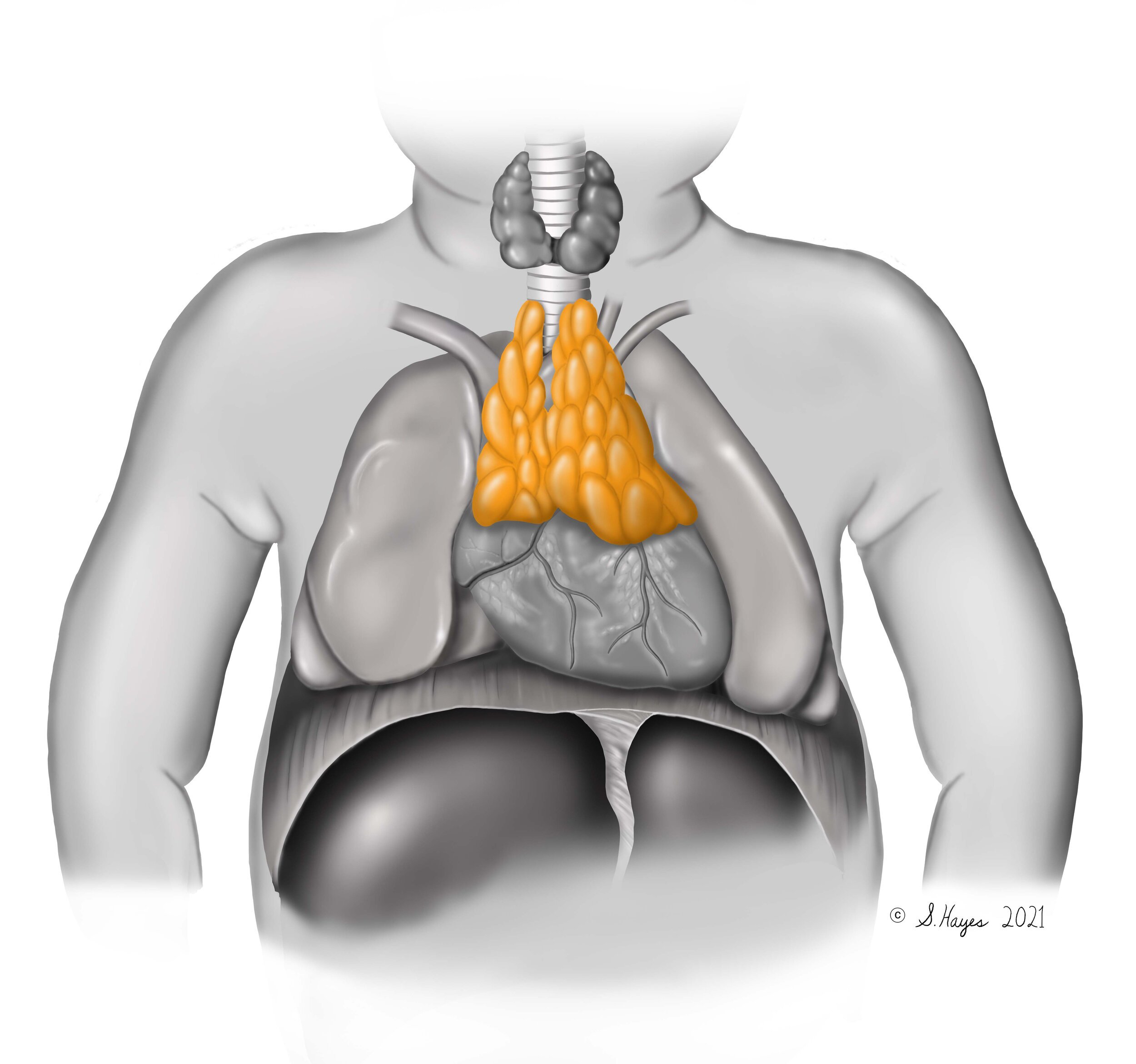
Thymus gland in an infant
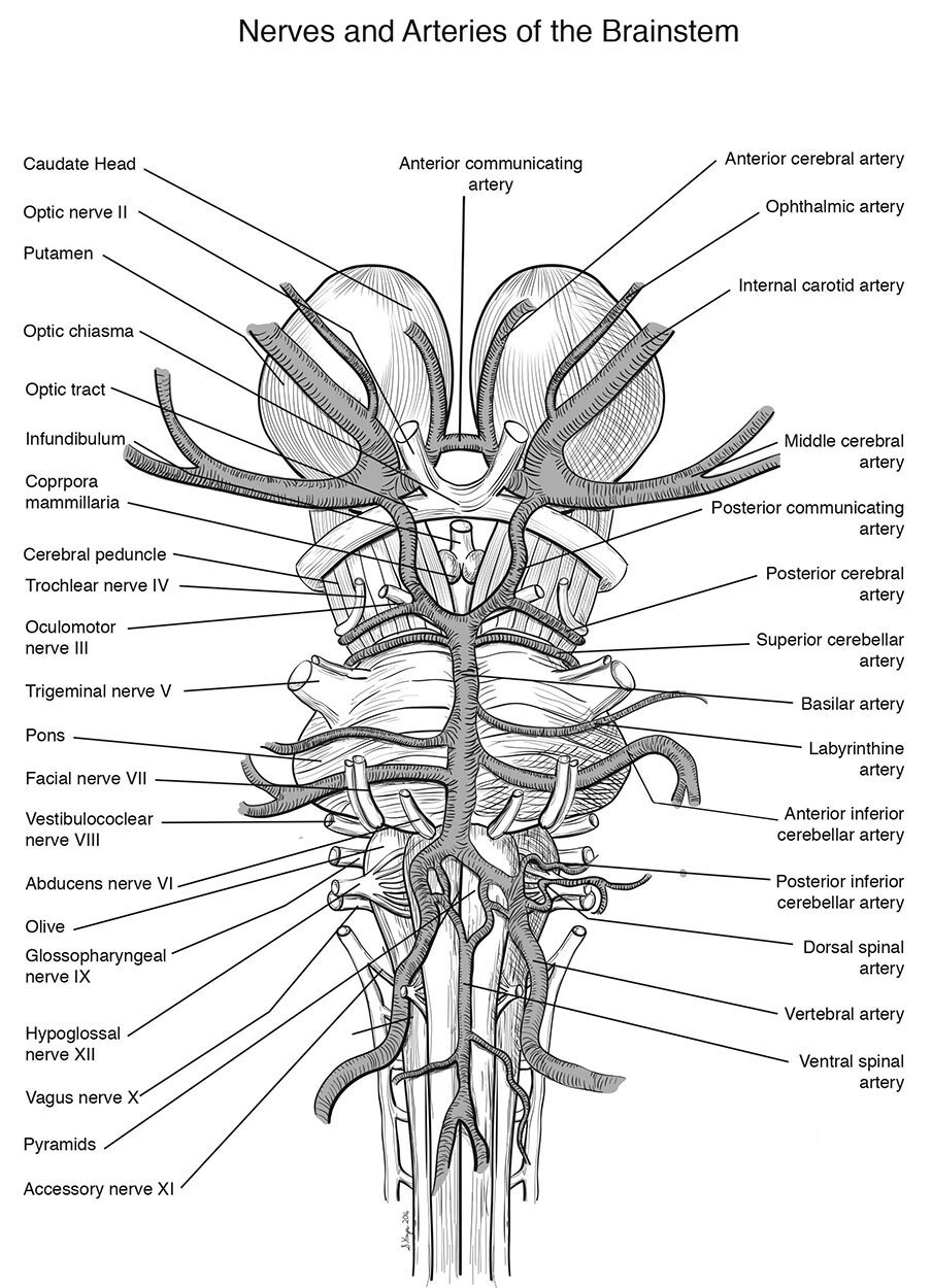
Nerves and Arteries of the Brainstem
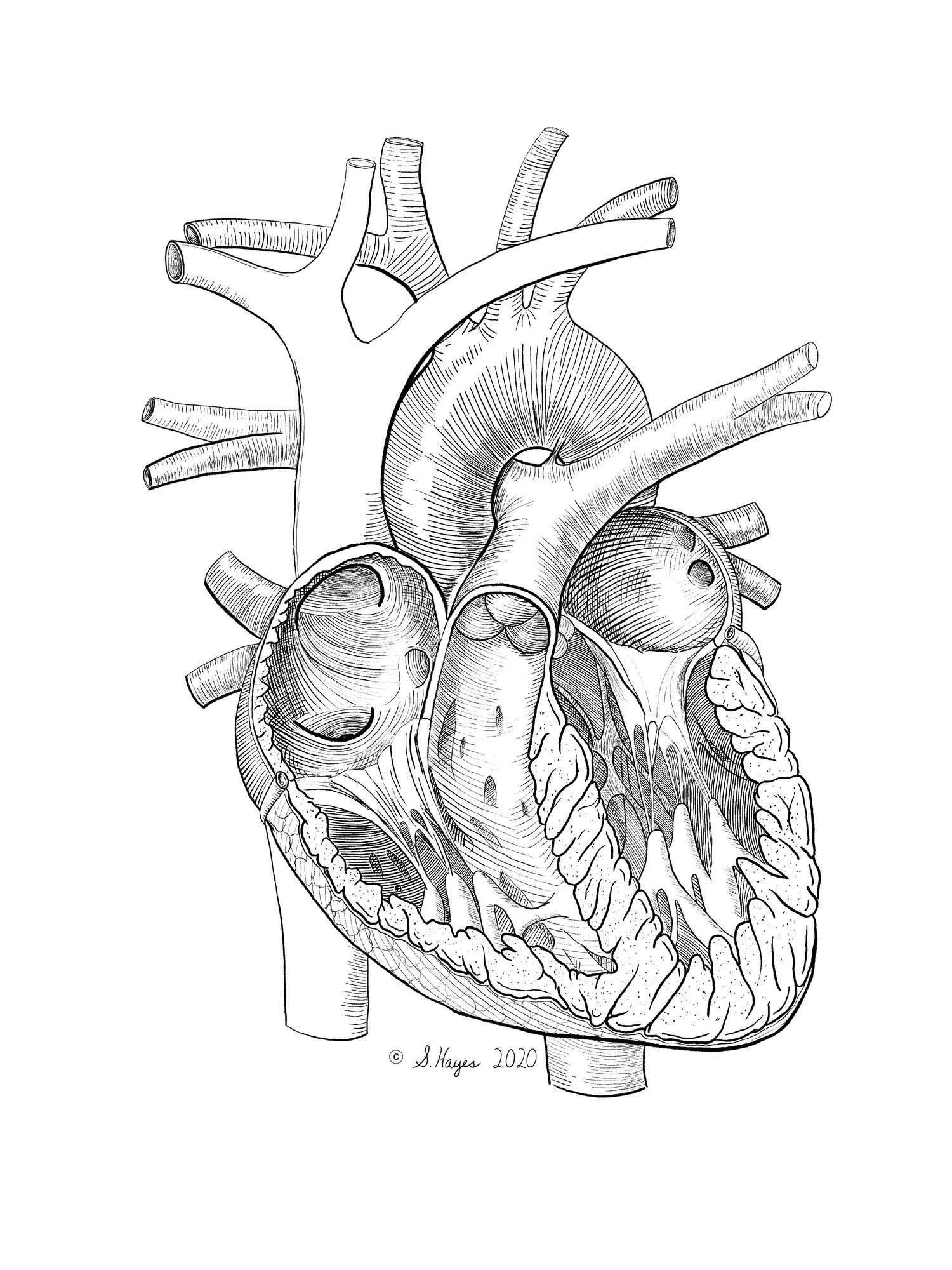
Heart

Ductal Carcinoma
Medical-Legal Exhibits
Medical-legal exhibits for trial, mediation, settlement, and promotional
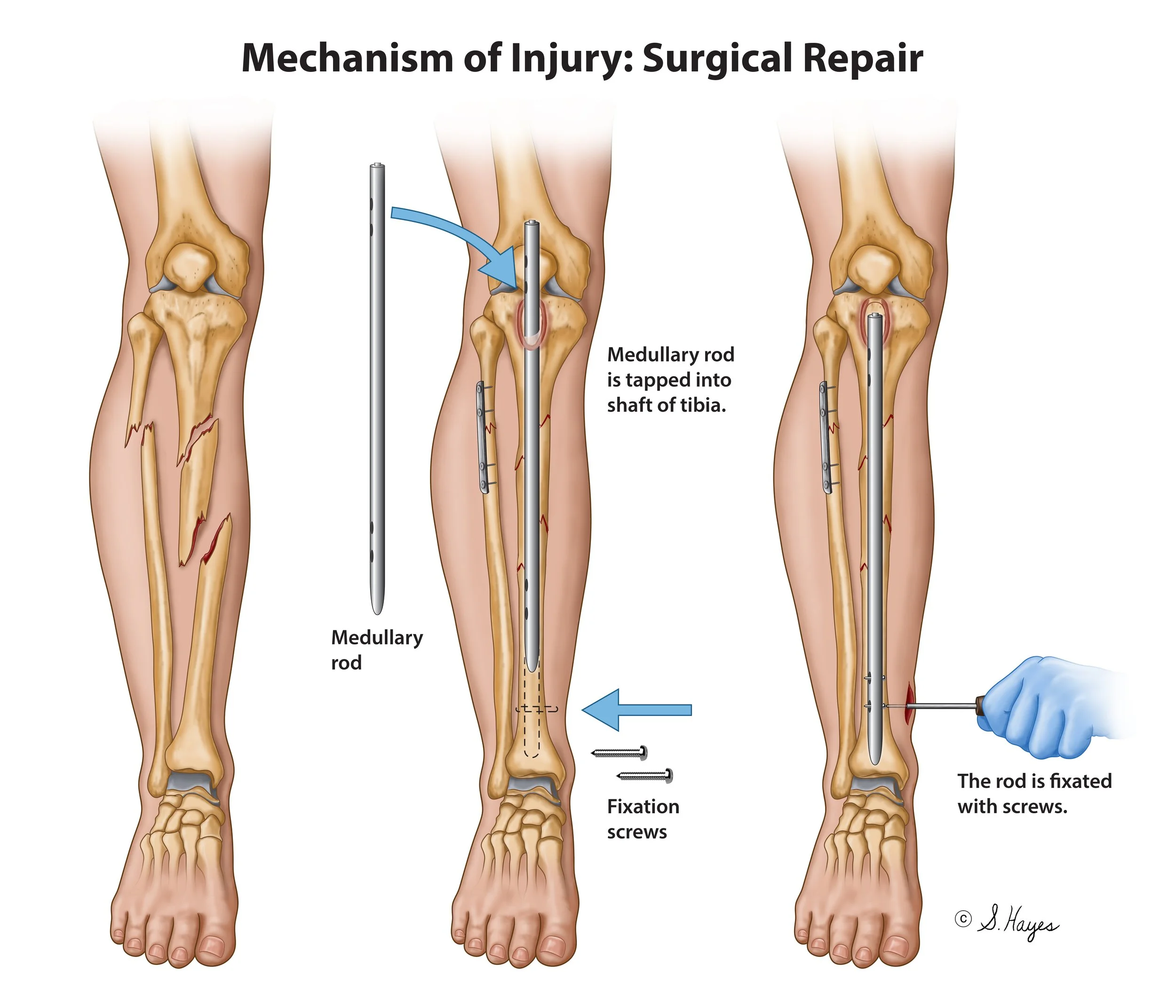
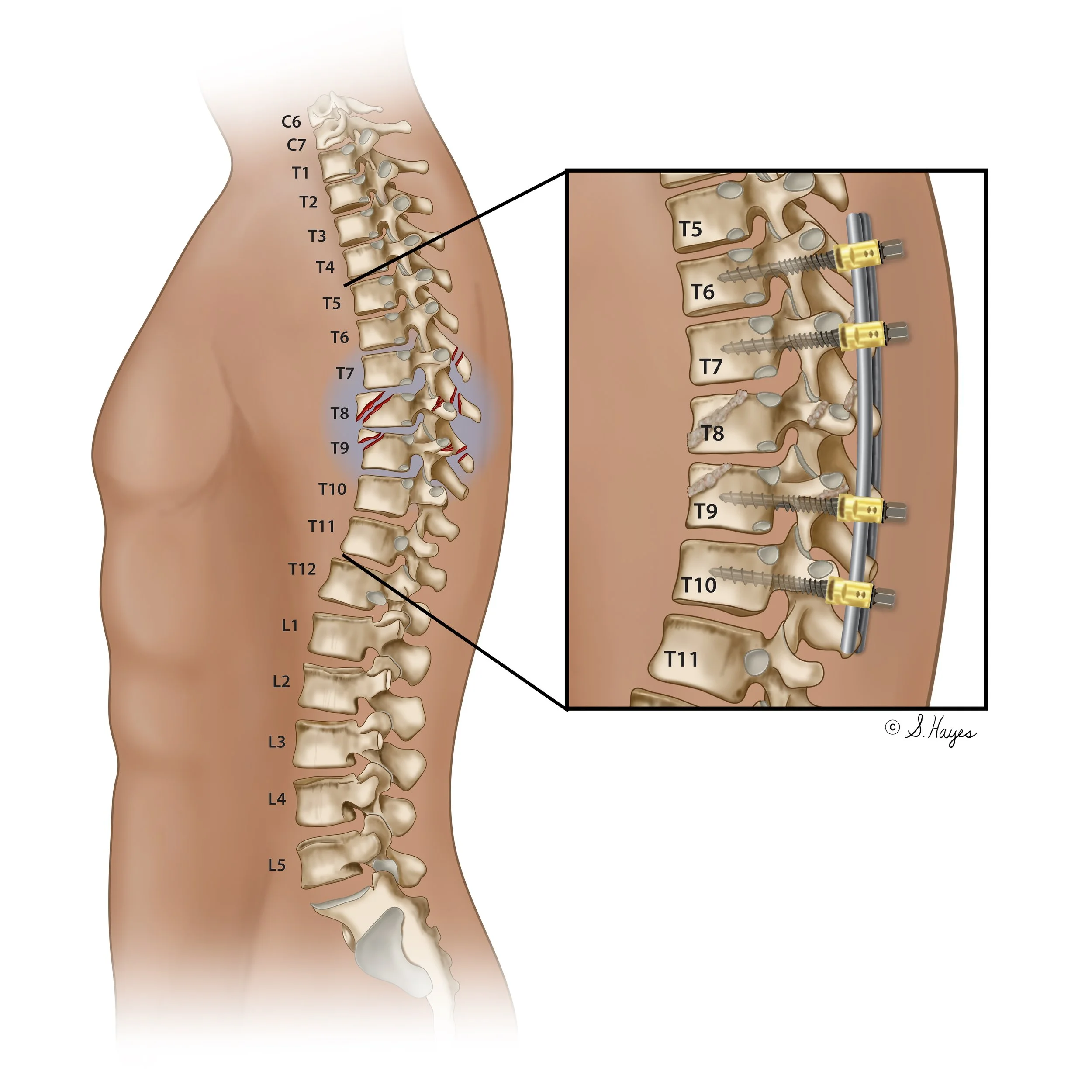
Mechanism of Injury: Surgical Repair
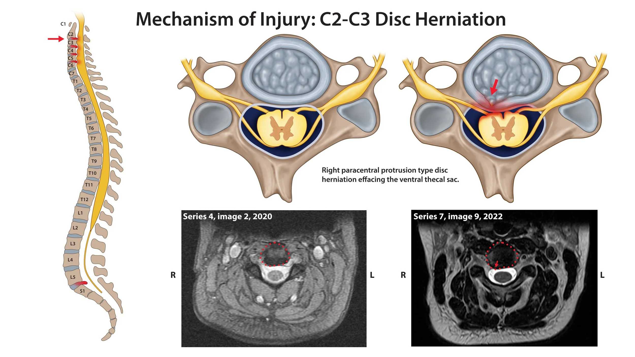
Mechanism of Injury: Disc Herniation

Long Bone Fracture Surgery
Crushed Vertebrate
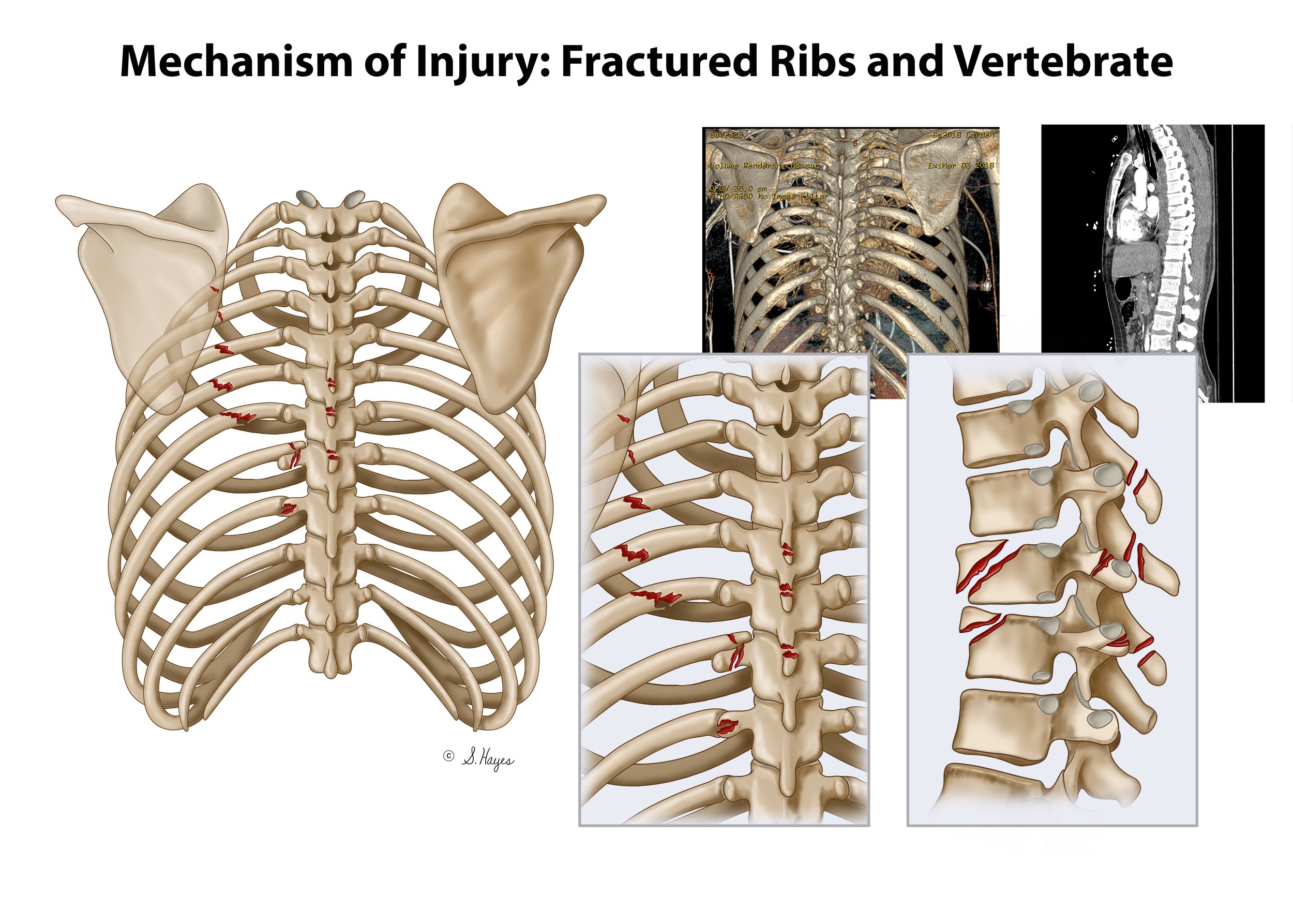
Diagram showing fractured ribs and vertebrae in the chest, with X-ray images in the background.
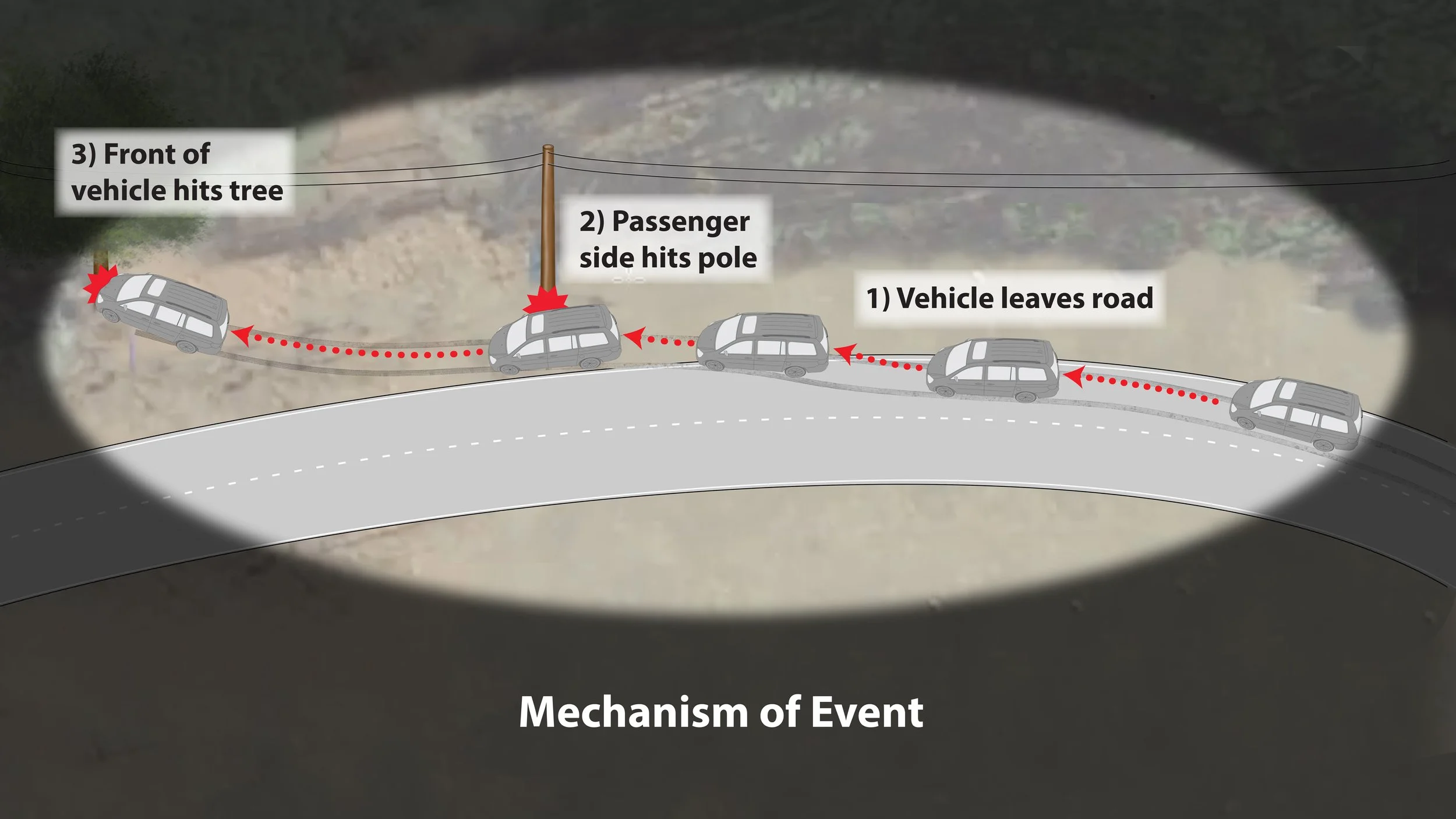
Diagram explaining the sequence of a vehicle hitting a tree. It shows a vehicle leaving the road, hitting a roadside pole, and then hitting a tree. Labels indicate the steps: 1) vehicle leaves road, 2) passenger side hits pole, 3) front of vehicle hits tree. The background is a rural area with electrical lines, and the image is viewed through a car's windshield.
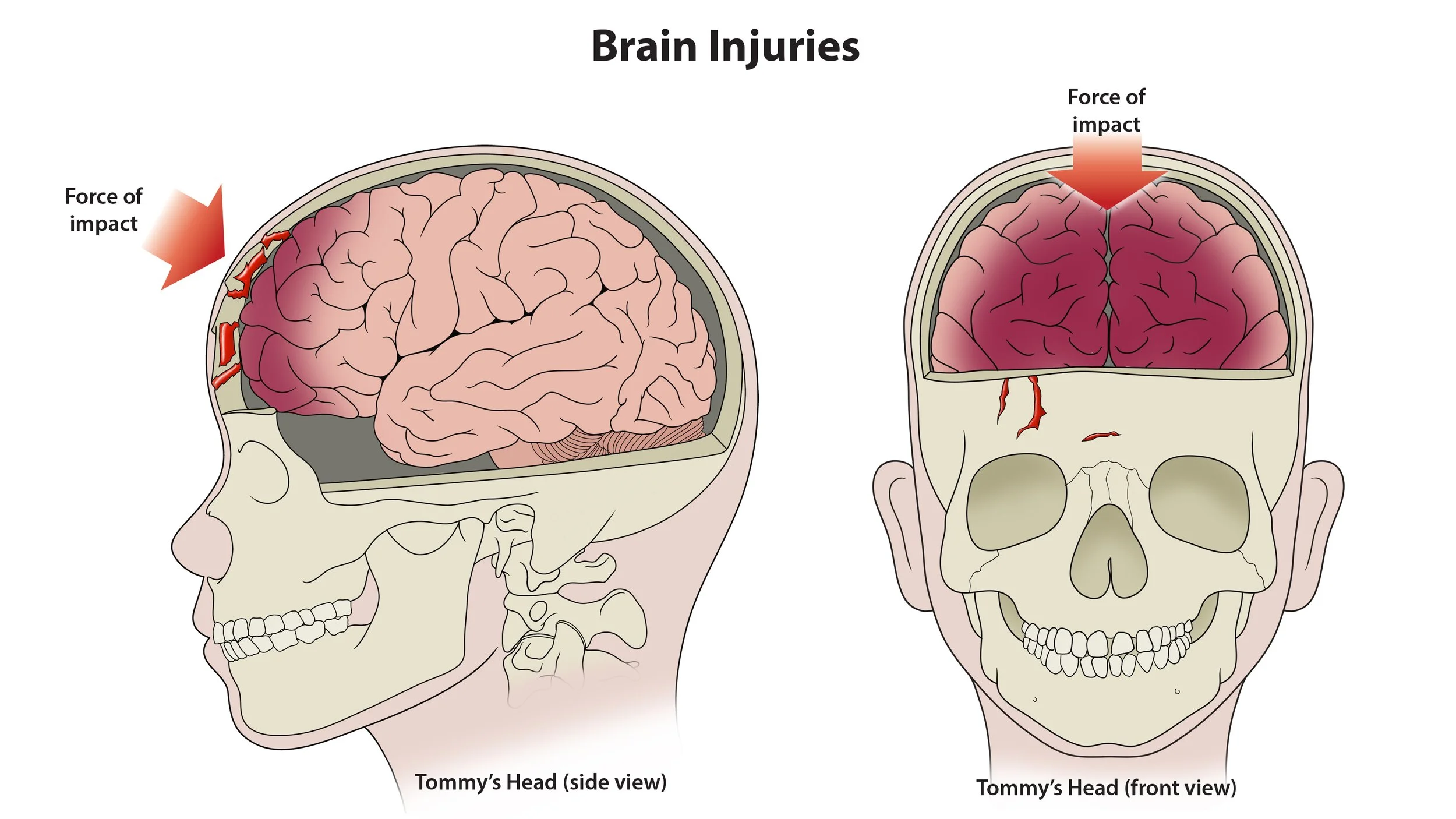
Diagram illustrating brain injuries from impact. Side and front views of a human head showing areas of brain damage and skull impact points.
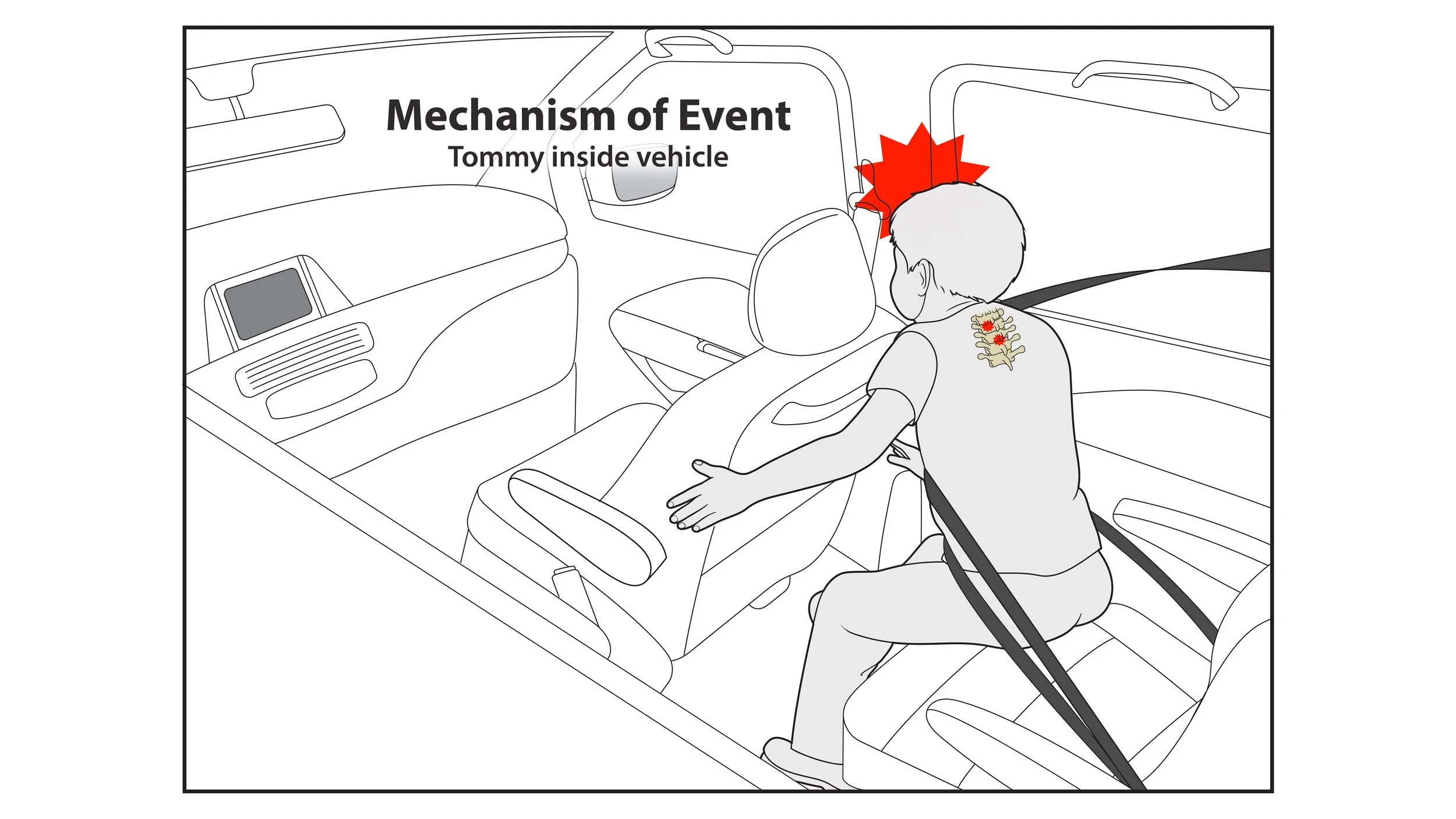
An illustration of a car interior showing a person hitting their head on the back of a front seat, with a red starburst indicating impact. The person is seated in the back seat and is wearing a shirt with a design on the back. The illustration explains the mechanism of a whiplash event caused by a rear-end collision.
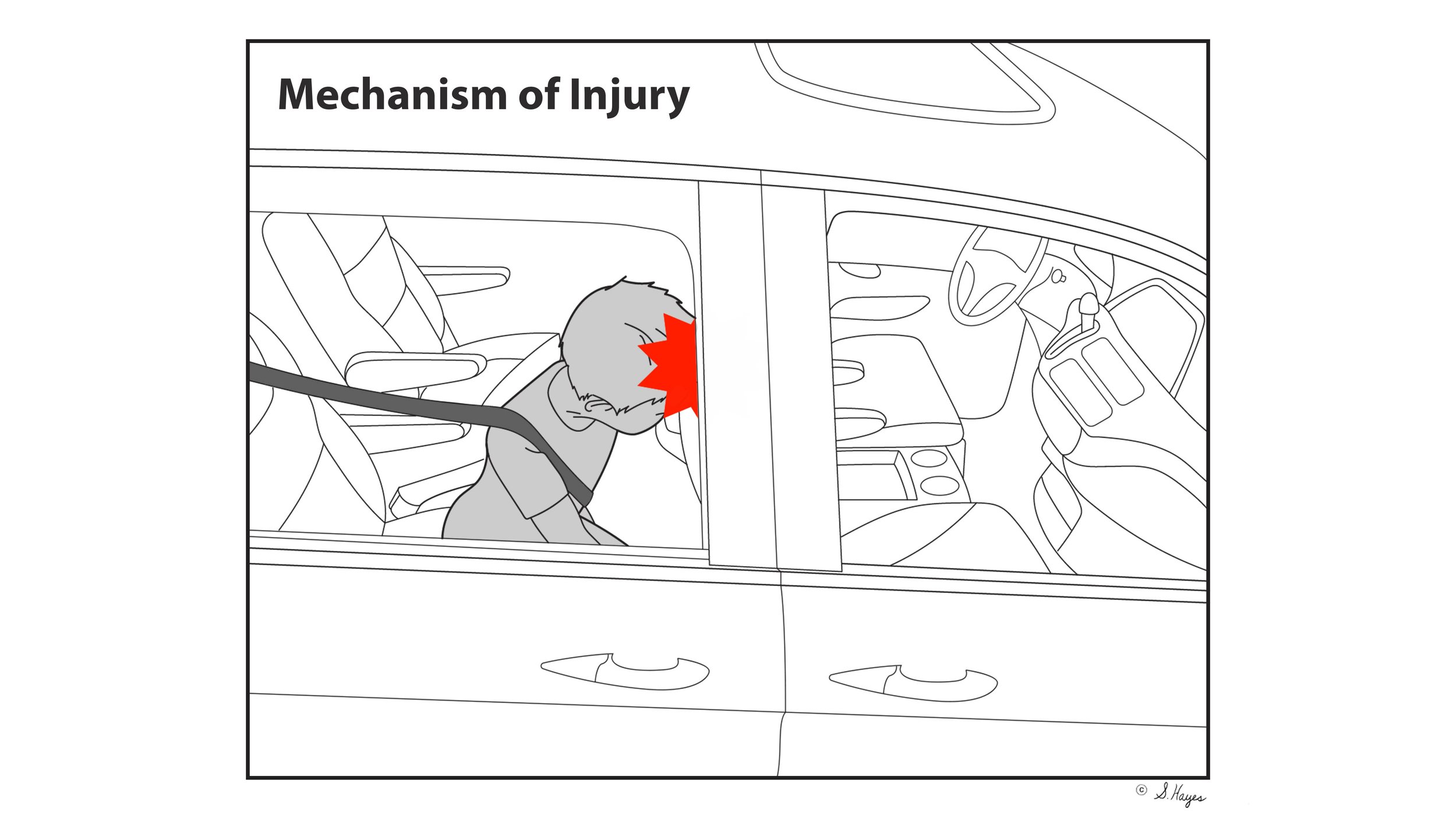
Diagram illustrating mechanism of injury with a person in a car, head hitting the front window, and a red explosion graphic indicating impact.
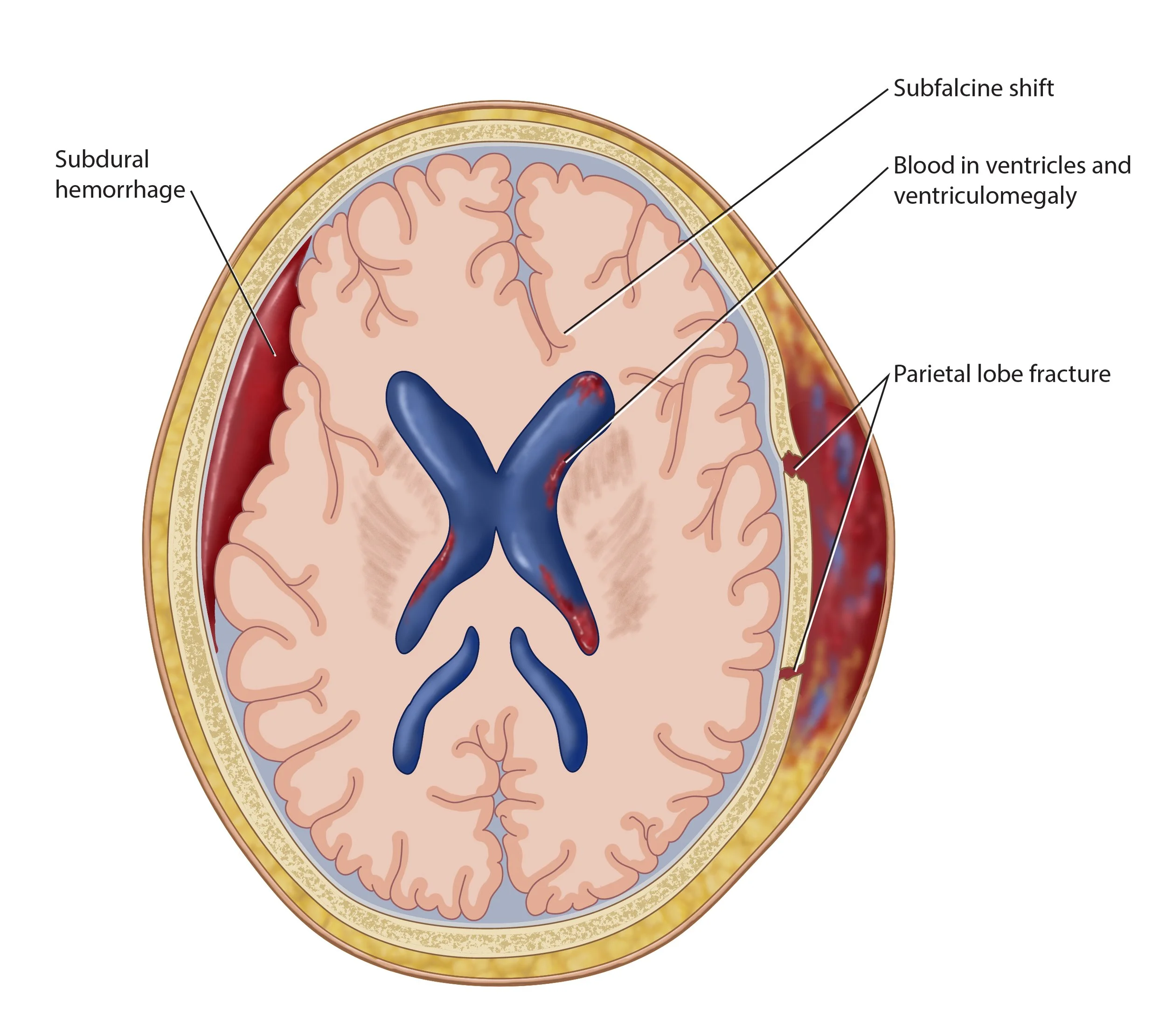
Subdural Hematoma
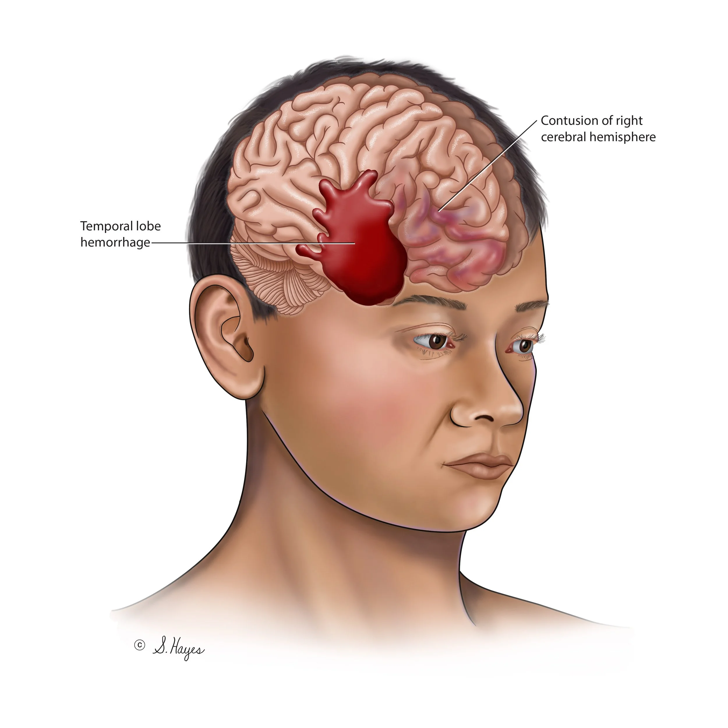
Illustration of a person with a cerebral hemorrhage, showing a bleeding in the temporal lobe and contusion of the right cerebral hemisphere.
3D Illustration
Models used to make medical illustration more dynamic, or for use in animation.
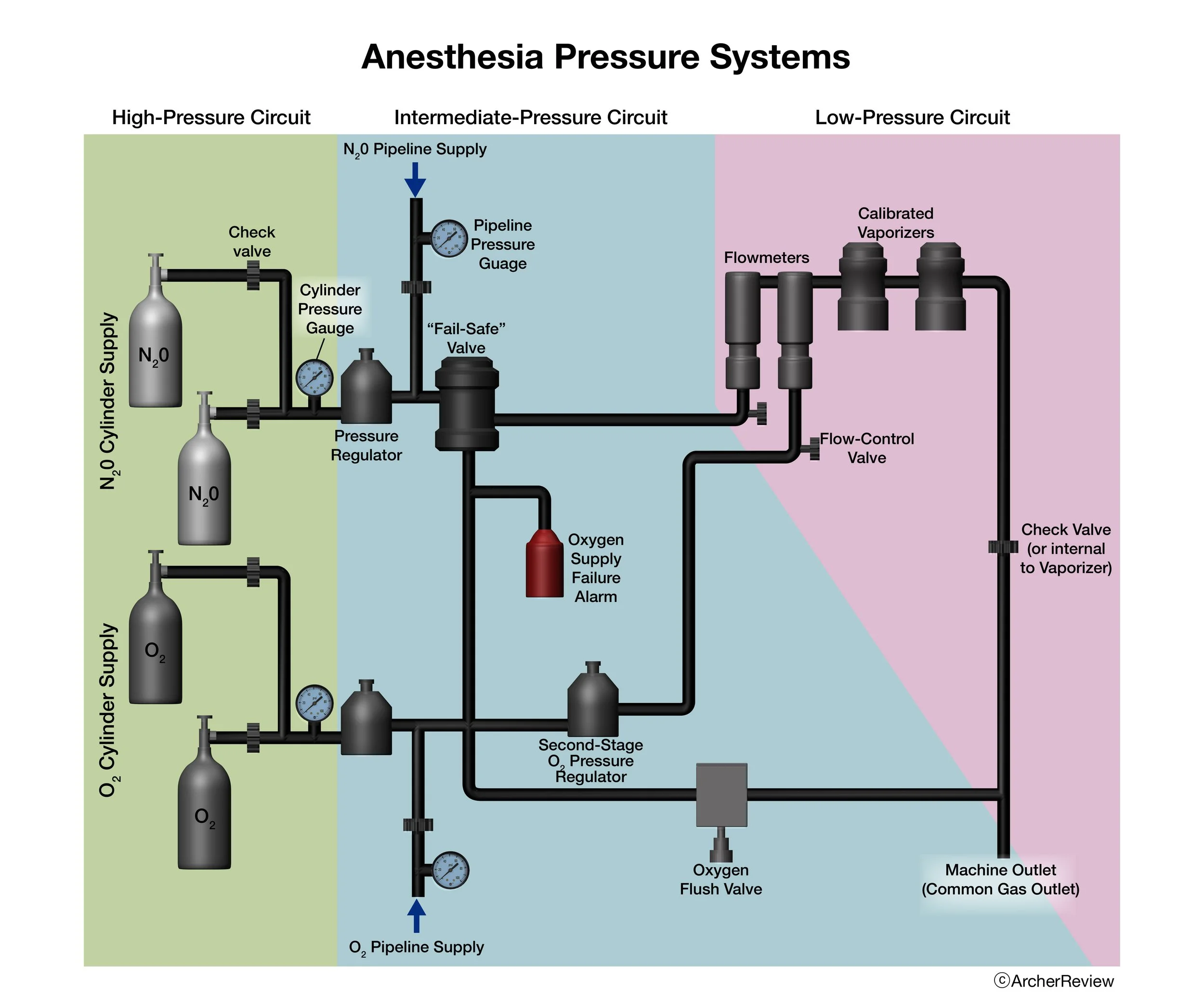
Anesthesia Pressure Systems

A digital illustration of DNA molecules with blue, green, orange, and cyan double helixes intertwined against a blue background with particles, representing genetic structure.

A digital illustration of DNA molecules, with green double helices connected by blue and orange strands, representing genetic material, against a background with a blue gradient and floating particles.
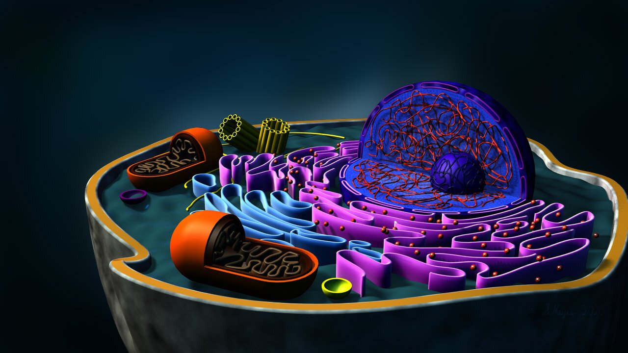
General Structure of an Animal Cell

Salmonella

Skin
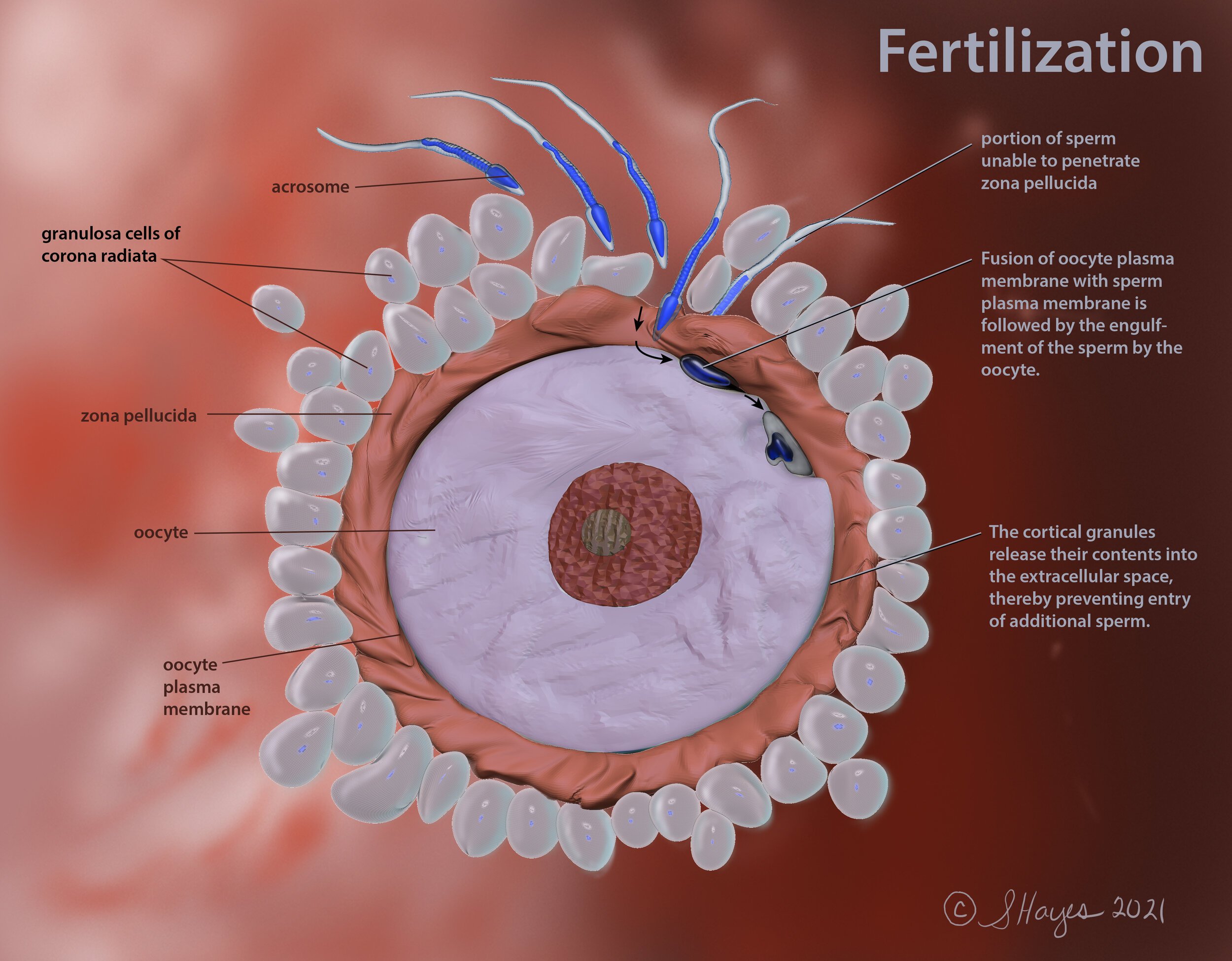
Fertilization of a Human Egg Cell
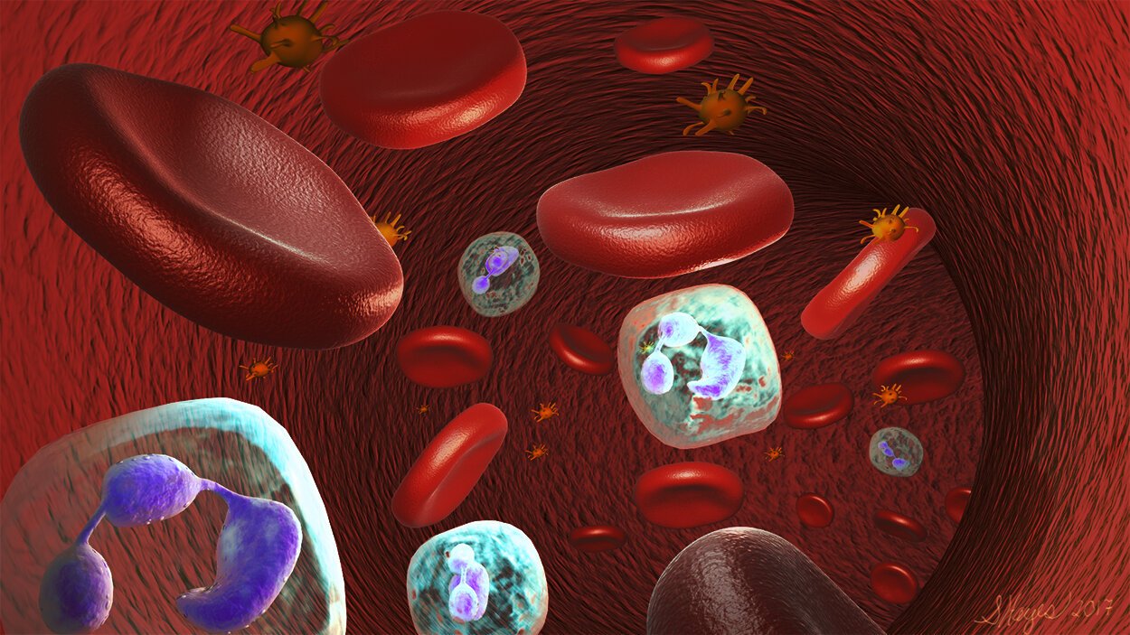
Cellular components of a blood vessel
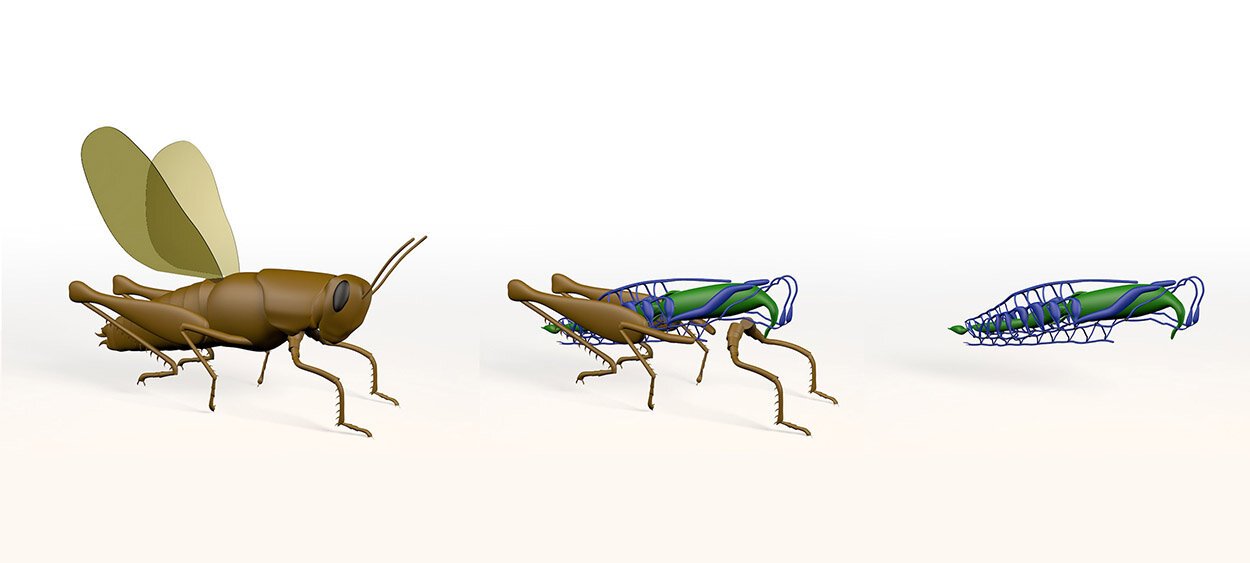
3D Grasshopper